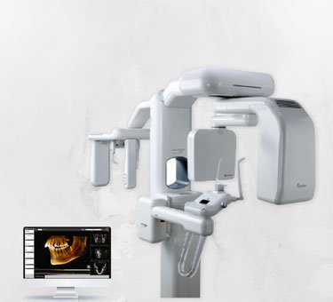

DentPrime offers you the best quality implants surgery in Turkey. You can also take advantage of free services such as online consultation, 3D tomography, volumetric tomography, free transfer, free hotel accommodations. Like hundreds of our patients, you will find us when you search such as best price of implants, dental implant packages, full mouth implant price, dental surgeon, dental surgery, best implantolog in Antalya, implant teeth, best quality dental treatments in Turkey, Specialist dentist in Turkey.
The use of imaging methods in the stages of diagnosis, treatment plan and treatment in oral and dental health will increase the success. For this purpose, two-dimensional imaging radiographs and three-dimensional tomographs can be used. With 3D imaging systems, jaw bones, cysts, impacted teeth, sinus cavities can be seen more clearly and accurately. In this way, more accurate and precise treatment planning can be made in all dental fields.
In DentPrime's clinic, only the best materials are used, and they are constantly inspected on an international level. The best result will come from a skilled dental surgeons with experience, a well-equipped dental lab, and skilled technicians.
With 3D tomography, all the measurements needed in the field of dentistry, surgery, implantology, periodontology, endodontics and orthodontics can be performed individually in three dimensions and increase the success of the treatment.
When dental tomographs work "volumetrically", it is also possible to obtain images with much lower doses than other tomographs.
With digital imaging systems, jaw bones, cysts, impacted teeth, sinus cavities can be seen more clearly and accurately. In this way, more accurate and precise treatment planning can be made in all dental fields.
At DentPrime Clinics, x-ray and 3D tomography services are offered to patients free of charge. Our aim is to determine the most appropriate treatment for our patients and increase their success rates.
Pre-implant treatment imaging is very important. Since three-dimensional imaging methods show the condition of the jaw bones very clearly, they contribute to the correct planning before starting the surgery. Information is obtained about the thickness, shape, interrelation of tissues and even bone density. Thus, before starting the procedure, it is determined which surgical techniques will be used and what can be done exactly. It greatly increases the success percentage of the transaction. After surgery, tomography can be taken under the control of implant placements and bone support.
With digital x-rays and 3d dental tomography, patients are exposed to less radiation than panoramic x-rays.
This is known as the background radiation we encounter in our daily life; Compared to radon gas, industrial waste, space and radiation from the earth, it is approximately equivalent to the amount of natural radiation of 7 hours per day for periapical radiographs and 2 days for panoramic radiographs. A 5-hour plane travel gives an amount of radiation that is almost equivalent to the radiation dose taken from a panoramic radiograph. The amount of radiation received by a person who smokes an average of 1 pack of cigarettes a day is equivalent to approximately 100 panoramic radiographs per year. Pregnant women and children should be more careful in all circumstances and lead vests should be used for all patients for protection purposes.
The use of imaging methods in the stages of diagnosis, treatment plan and treatment in oral and dental health will increase the success. For this purpose, two-dimensional imaging radiographs and three-dimensional tomographs can be used. With 3D imaging systems, jaw bones, cysts, impacted teeth, sinus cavities can be seen more clearly and accurately. In this way, more accurate and precise treatment planning can be made in all dental fields.
With 3D tomography, all the measurements needed in the field of dentistry, surgery, implantology, periodontology, endodontics and orthodontics can be performed individually in three dimensions and increase the success of the treatment. When dental tomographs work "volumetrically", it is also possible to obtain images with much lower doses than other tomographs. With digital imaging systems, jaw bones, cysts, impacted teeth, sinus cavities can be seen more clearly and accurately. In this way, more accurate and precise treatment planning can be made in all dental fields.
Pre-implant treatment imaging is very important. Since three-dimensional imaging methods show the condition of the jaw bones very clearly, they contribute to the correct planning before starting the surgery. Information is obtained about the thickness, shape, interrelation of tissues and even bone density. Thus, before starting the procedure, it is determined which surgical techniques will be used and what can be done exactly. It greatly increases the success percentage of the transaction. After surgery, tomography can be taken under the control of implant placements and bone support.
With digital x-rays and 3-dimensional dental tomography, patients are exposed to less radiation than panoramic x-rays.
This is known as the background radiation we encounter in our daily life; Compared to radon gas, industrial waste, space and radiation from the earth, it is approximately equivalent to the amount of natural radiation of 7 hours per day for periapical radiographs and 2 days for panoramic radiographs.
A 5-hour plane travel gives an amount of radiation that is almost equivalent to the radiation dose taken from a panoramic radiograph.
The amount of radiation received by a person who smokes an average of 1 pack of cigarettes a day is equivalent to approximately 100 panoramic radiographs per year.
Pregnant women and children should be more careful in all circumstances and lead vests should be used for all patients for protection purposes.
• Detection of caries
• Before and during root canal treatment
• Detecting bone damage in cases with advanced gum disease
• Pre-implant surgery planning and follow-up of treatment
• Joint disorders,
• In case of cysts and tumors related to teeth and bones
• Determining the position of the impacted tooth before surgical procedures
• In orthodontic treatment, in determining the relationship between the jaws and teeth
• Salivary gland diseases
• Following the tooth development and growth of children
• In suspicion of tooth and jaw fracture