





Undergoing a surgical or simple dental extraction initiates a complex biological cascade inside the oral cavity. As the body begins to heal the socket, many patients run their tongue across the surgical site a few days or weeks later and feel a sharp, hard projection poking through the soft tissue. Experiencing a bone spur after tooth extraction is a common occurrence during alveolar socket healing. While finding a sharp fragment often causes patients to worry that a broken tooth root was left behind, these occurrences represent natural anatomical responses of the jawbone during bone remodeling.
Patients experiencing post-operative changes frequently ask what is a bone spur after tooth extraction and seek reassurance regarding oral safety. Learning what causes bone spur after tooth extraction helps distinguish natural physiological bone sequestration from pathological complications. Whether a patient discovers a large bone spur after tooth extraction, suffers when a bone spur after tooth extraction painful episode irritates the lateral borders of the tongue, or notices an exposed gum bone spur after tooth extraction, this clinical guide provides comprehensive answers for safe oral recovery.
Table Of Contents
- What Is a Bone Spur After Tooth Extraction and How Does It Form?
- Etiology: What Causes Bone Spur After Tooth Extraction?
- Clinical Symptoms: When Is a Bone Spur After Tooth Extraction Painful?
- Differential Diagnosis: Bone Spur vs. Retained Tooth Root Tip
- Treatment and Removal Protocols: How Dentists Manage Bone Spurs
- Comprehensive Comparison: Healing Timelines and Bone Behaviors
- Frequently Asked Questions
- What is a bone spur after tooth extraction?
- What causes bone spur after tooth extraction to develop?
- How do I know if my bone spur after tooth extraction is painful or infected?
- Can a large bone spur after tooth extraction go away on its own?
- Should I try to remove a gum bone spur after tooth extraction at home?
What Is a Bone Spur After Tooth Extraction and How Does It Form?
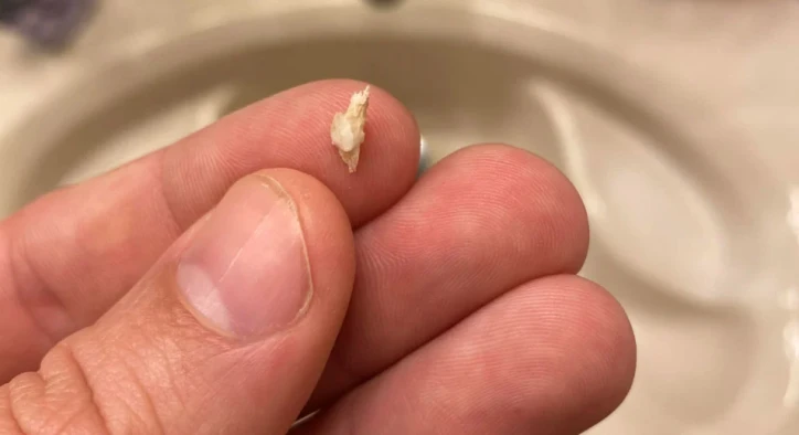
When examining socket healing, patients naturally ask: what is a bone spur after tooth extraction? Clinically known as an osteophyte, bone spicule, or bone sequestrum, it is a small, sharp fragment of bone that works its way through the gum tissue after a tooth has been pulled. Teeth sit anchored within bony sockets called alveoli. The bone walls surrounding the roots are extremely thin, especially on the outer facial and inner lingual plates.
- [ TOOTH EXTRACTION COMPLETED ]: Thin alveolar socket walls lose mechanical tooth support
- [ SPICULE EXTERIORIZATION ]: Sharp fragment exfoliates through mucosa or resorbs naturally
When a tooth is removed, the surrounding alveolar bone loses its blood supply from the periodontal ligament. The body initiates remodeling through specialized cells called osteoclasts, which resorb unnecessary bone margins, while osteoblasts lay down new bone inside the socket. During this restructuring, tiny pieces of the delicate socket wall can break off or become non-vital. As the healing gum tissue contracts and matures, it pushes these sharp bony fragments outward toward the oral cavity, leading to the development of a noticeable gum bone spur after tooth extraction.
Etiology: What Causes Bone Spur After Tooth Extraction?
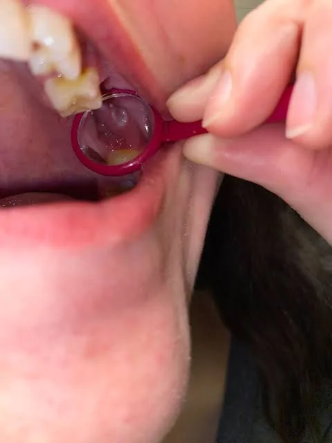
Patients recovering from dental surgery routinely ask what causes bone spur after tooth extraction. Bone spicules do not result from surgical error; rather, they arise from natural anatomical and mechanical factors during tooth removal.
- Micro-Fractures of the Alveolar Crest: Teeth with curved roots, dense bone, or ankylosis require controlled lateral pressure during elevation. This mechanical pressure can cause micro-fractures in the razor-thin crestal bone of the socket rim.
- Bone Remodeling Dynamics: After tooth removal, the alveolar ridge undergoes significant vertical and horizontal volume reduction. Sharp underlying bone edges that are not resorbed quickly by osteoclasts end up protruding through thinning mucosal tissue.
- Surgical Tooth Sectioning: Impacted wisdom teeth or multi-rooted molars often require sectioning with surgical burs. Microscopic bone chips produced during tooth separation can remain within socket crevices and surface weeks later.
- Pre-Existing Periapical Infection: Chronic dental infections cause localized bone inflammation. The compromised blood flow in infected areas can leave tiny areas of dead cortical bone (sequestra) that the body systematically rejects.
These factors demonstrate that whether dealing with a minor spicule or a large bone spur after tooth extraction, the condition reflects the body's natural effort to expel non-vital bone fragments.
Clinical Symptoms: When Is a Bone Spur After Tooth Extraction Painful?
A bone spicule can range from an unnoticeable rough spot to an uncomfortable complication. Understanding why a bone spur after tooth extraction painful condition develops helps patients manage discomfort effectively.
- [ SHARP BONE PROTRUSION ]: Penetrates mucosal barrier, causing mechanical friction
- [ CLINICAL RESOLUTION ]: Gentle extraction by dentist or spontaneous physiological exfoliation
The sharp bone edge itself contains no nerve endings once separated from the periosteum. However, as the jagged edge pierces through the sensitive, richly innervated gum tissue, it causes localized inflammation. Furthermore, constant friction from the tongue rubbing against the sharp edge creates painful aphthous-like ulcers. Patients often experience soreness during chewing, speaking, or swallowing, especially when a spicule forms on the lower jaw near the mylohyoid ridge.
Differential Diagnosis: Bone Spur vs. Retained Tooth Root Tip
One of the most frequent clinical concerns expressed by patients is confusing a bone fragment with a retained tooth root. Understanding their distinct diagnostic traits brings peace of mind.
Diagnostic Feature | Bone Spur (Bone Spicule) | Retained Tooth Root Tip |
Tissue Origin | Alveolar jawbone (cortical bone) | Tooth structure (dentin and enamel) |
Color Appearance | Chalky white, yellowish, or creamy bone tone | Creamy white, translucent, or dark pulp outline |
Mobility | Often slightly loose and wiggles when nudged | Firmly wedged and immovable in jawbone |
Radiographic Appearance | Irregular, thin radiopaque outline with low density | Distinct root canal shape with periodontal ligament |
Clinical Resolution | Naturally resorbs or exfoliates over 2 to 6 weeks | Requires clinical monitoring or surgical retrieval |
Treatment and Removal Protocols: How Dentists Manage Bone Spurs
Discovering a large bone spur after tooth extraction does not always require immediate surgery. Dentists choose between conservative monitoring and gentle chairside intervention based on clinical severity.
- [ MINOR ASYMPTOMATIC SPICULE ]: Warm saltwater rinses & observation (Resorbs in 2 to 4 weeks)
- [ IN-OFFICE ALVEOLOPLASTY ]: Local anesthesia, gentle bone smoothing with rongeur or bur
- Conservative Observation: Many small spicules work their way completely out of the gum tissue on their own within two to four weeks. During this time, the body either dissolves the fragment with osteoclasts or pushes it free into the mouth where it sheds harmlessly.
- Warm Saltwater Rinses: Rinsing with half a teaspoon of salt dissolved in warm water four times daily reduces localized gum swelling, prevents superficial bacterial infection, and accelerates soft tissue healing.
- Chairside Alveoloplasty: If a bone spur causes severe pain, fails to shed, or presents as a prominent ridge that lacerates the tongue, a dentist performs a simple chairside smoothing procedure. Under local anesthesia, the clinician uses a bone file, rongeur forceps, or a diamond bur to smooth down the sharp projection in minutes.
- Never Pick at the Fragment: Patients must never use tweezers, needles, or fingernails to pry out a bone spur at home. Unsterile tools introduce bacteria deep into healing alveolar bone, which can cause severe alveolar osteitis (dry socket) or widespread bone infection.
Comprehensive Comparison: Healing Timelines and Bone Behaviors
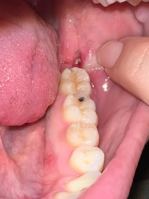
Evaluating the timeline of post-extraction alveolar bone healing illustrates why bone spurs appear weeks after surgery.
Healing Stage | Post-Extraction Timeline | Biological Bone Behavior |
Hemostasis & Clotting | Hours 1 to 24 | Blood clot fills socket; bone edges exposed internally |
Granulation Ingrowth | Days 3 to 7 | Soft granulation tissue covers base; bone crest begins remodeling |
Spicule Emergence | Weeks 2 to 4 | Remodeling pushes non-vital bone fragments toward gum surface |
Trabecular Woven Bone | Weeks 6 to 8 | Immature woven bone fills socket; surface bone rounds out |
Lamellar Bone Maturation | Months 3 to 6 | Fully corticalized alveolar ridge ready for implants or bridges |
Frequently Asked Questions
What is a bone spur after tooth extraction?
A bone spur after tooth extraction is a small, sharp piece of dead or non-vital alveolar bone that surfaces through the gums as the extraction socket heals and remodels. It is not a piece of a tooth root, but rather a normal fragment of the bone that used to support the tooth.
What causes bone spur after tooth extraction to develop?
Bone spurs develop due to mechanical micro-fractures occurring during tooth elevation, rapid resorption of thin alveolar socket walls, and the natural remodeling process where the body pushes out non-vital bone fragments as the gums shrink and heal over the socket.
How do I know if my bone spur after tooth extraction is painful or infected?
A bone spur causes localized pain when its sharp edges poke through the gums or rub against the tongue, creating small ulcers. However, if you experience throbbing jaw pain, persistent foul odor, foul taste, facial swelling, or fever, you should see your dentist promptly to rule out dry socket or localized bone infection.
Can a large bone spur after tooth extraction go away on its own?
Yes. Many bone spurs, even relatively large ones, naturally loosen and fall out on their own as the gums heal, or the body slowly resorbs them over two to six weeks. However, if a large spur causes constant pain or prevents normal eating, your dentist can smooth or remove it in minutes.
Should I try to remove a gum bone spur after tooth extraction at home?
No. You should never attempt to pick, pull, or cut out a bone spur using household tweezers, pins, or fingernails. Prying at the bone damages healing gums, exposes deeper bone structures to oral bacteria, and risks serious infections or dry socket complications.
About the Author