





Adapting to a removable complete or partial prosthetic appliance represents a substantial physiological transition for the oral tissues. In clinical prosthodontics, the appearance of denture sores is one of the most common early complications reported by patients following delivery or long-term wear. Rather than indicating an intrinsic failure of prosthetic treatment, these localized erosive lesions highlight localized pressure concentrations where rigid acrylic resin impinges upon the delicate, non-keratinized alveolar mucosa, producing painful mechanical ulcerations or chronic fungal inflammation beneath the denture base.
Patients struggling with everyday chewing discomfort frequently report a tender, painful sore gum from dentures and wonder whether feeling sore from dentures during the initial weeks is biologically expected. Many search for practical home recommendations regarding what to use for denture sores to ease burning sensations, while others ask with urgency what is the fastest way to heal denture sores before attending an adjustment appointment. Furthermore, distinguishing between localized pressure decubitus ulcers and generalized, bright red denture stomatitis is essential for determining whether an antifungal denture stomatitis treatment or an in-office acrylic modification is clinically required. This clinical guide delivers transparent medical facts to restore comfort and tissue health.
Table Of Contents
- Biomechanical Architecture: What Causes Denture Sores?
- Etiology: Identifying Primary Drivers of Mucosal Trauma
- Fungal Infections: Denture Stomatitis and Targeted Treatment
- Emergency Relief: What to Use for Denture Sores at Home
- Accelerated Recovery: What Is the Fastest Way to Heal Denture Sores?
- Comprehensive Comparison: Traumatic Denture Ulcers vs. Denture Stomatitis
- Frequently Asked Questions
- How do I know if my sore gum from dentures is normal or an infection?
- What is the fastest way to heal denture sores without ruining my denture?
- What to use for denture sores to relieve pain while eating?
- Can denture stomatitis spread to other parts of my body?
- Why do I suddenly have a sore from dentures that I have worn for years?
Biomechanical Architecture: What Causes Denture Sores?
To understand why mucosal ulceration occurs, one must examine the histological differences between periodontal ligaments and edentulous ridge mucosa. Natural teeth distribute chewing forces through resilient collagen fibers embedded directly into the alveolar bone. In contrast, removable dentures rest directly on oral mucosa that is merely two to three millimeters thick.
- [ ACRYLIC OVER-EXTENSION OR IRREGULARITY ]: Uneven bite force focuses on a single bone ridge
- [ MECHANICAL DECUBITUS ULCER ]: Painful, raw sore with erythematous border develops
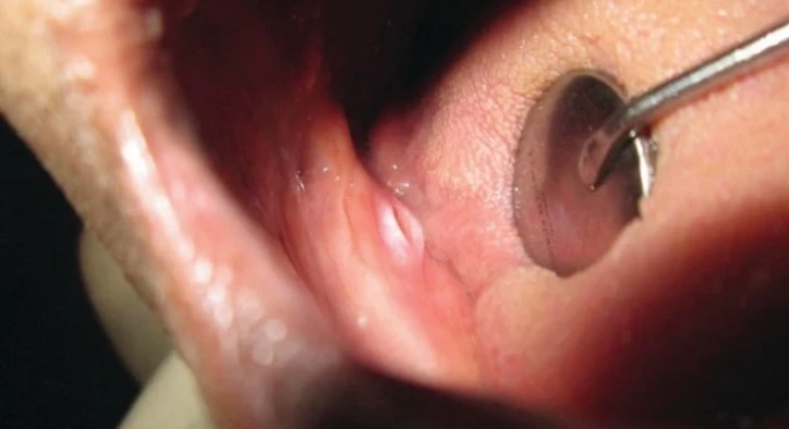
When an acrylic flange extends too deeply into the labial or lingual sulcus, or when uneven bite contacts tilt the denture base during mastication, the underlying capillary network is compressed against the hard cortical bone. This localized ischemia starves the epithelial cells of oxygen and nutrients, leading to cellular breakdown and sloughing within twenty-four to forty-eight hours. The resulting mucosal erosion presents clinically as a sharp, exquisitely tender ulcer surrounded by an inflamed red halo, making continued denture wear excruciating.
Identifying Primary Drivers of Mucosal Trauma
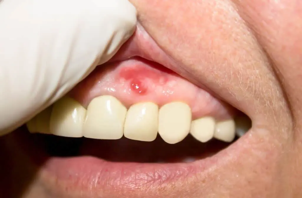
A persistent sore rarely emerges without specific physical, biological, or anatomical catalysts acting on the denture-bearing foundation.
- Over-Extended Flanges: When border molding or impression trays capture excess depth in the vestibular sulcus, the rigid acrylic edge continuously digs into movable muscular attachments during speech and swallowing.
- Occlusal High Spots: If upper and lower artificial teeth do not interdigitate evenly, a premature contact on one side drives the opposite or adjacent denture base violently into the alveolar ridge with every chewing cycle.
- Alveolar Bone Resorption: Over time, the edentulous jawbone naturally shrinks and recedes, causing a once-perfectly fitting denture to loosen, rock, and slide, generating friction-induced blister points.
- Rough Acrylic Imperfections: Microscopic nodules or processing flaws on the tissue-fitting surface of newly cured polymethyl methacrylate scratch delicate mucosal ridges.
- Continuous Day and Night Wear: Wearing dentures twenty-four hours a day deprives the underlying mucosa of salivary flow and atmospheric oxygen, drastically reducing tissue recovery and promoting ulceration.
Fungal Infections: Denture Stomatitis and Targeted Treatment
When mucosal redness is generalized beneath the entire upper denture base rather than isolated to a localized pressure spot, clinicians diagnose denture stomatitis.
- [ CONTINUOUS OCCLUSION & DRYNESS ]: Acrylic surface accumulates thick fungal biofilm
- [ ERYTHEMATOUS STOMATITIS ]: Generalized bright-red mucosal inflammation without ulceration
Classified under Newton's criteria from pinpoint hyperemic lesions (Type One) to generalized velvety erythema (Type Two) and papillary hyperplasia (Type Three), this condition is primarily driven by Candida albicans colonizing the microporous acrylic fitting surface. Unlike localized traumatic ulcers, stomatitis often causes minimal acute pain, presenting instead with mild burning, dry mouth, or altered taste.
A definitive denture stomatitis treatment protocol requires treating both the oral mucosa and the acrylic appliance simultaneously. The dentist prescribes topical antifungal miconazole gel or nystatin oral suspensions for the tissue, paired with daily soaking of the denture in diluted sodium hypochlorite solutions or chlorhexidine gluconate to sterilize the porous resin. If the fungal reservoir on the prosthesis is ignored, the oral infection will recur immediately upon reinsertion.
Emergency Relief: What to Use for Denture Sores at Home
When patients seek guidance on what to use for denture sores to ease daily eating, practical over-the-counter and home measures provide symptomatic relief before clinical chairside adjustments.
[ MUCOSAL SORE IDENTIFIED ]: Remove appliance to relieve pressure; warm salt water rinse
[ PROSTHODONTIC ADJUSTMENT ]: Pressure-indicator paste mapping and acrylic relief
- Warm Salt Water Rinses: Dissolve half a teaspoon of salt in a glass of warm water and gently rinse the mouth for thirty to sixty seconds three to four times daily. Hypertonic saline reduces intercellular edema, soothes exposed nerve endings, and creates an inhospitable environment for pathogenic bacteria.
- Topical Anesthetic Barrier Gels: Formulations containing twenty percent benzocaine or soothing hyaluronic acid temporarily numb superficial pain receptors while creating a protective bio-adhesive shield over raw ulcer surfaces.
- Rest the Oral Tissues: The single most crucial step is removing the denture for several hours during the day and never sleeping with it in place. Tissue healing cannot initiate if mechanical friction continuously rubs the open wound.
- Never File the Denture Yourself: Do not attempt to adjust, sand, or file down the acrylic appliance at home using nail files, knives, or sandpaper. Inaccurate home trimming alters delicate bite geometry, ruins retention, and creates rough surfaces that generate worse secondary ulcers.
Accelerated Recovery: What Is the Fastest Way to Heal Denture Sores?
Patients experiencing debilitating pain routinely ask: what is the fastest way to heal denture sores? Clinically, the fastest protocol relies on immediate professional pressure relief combined with tissue conditioning.
The healing timeline accelerates dramatically once a prosthodontist uses Pressure Indicator Paste (PIP) to map the exact internal interference. By painting a thin layer of white zinc oxide paste inside the denture and seating it in the mouth, high-pressure areas displace the paste, showing through clearly. The dentist selectively relieves the offending acrylic spot with a carbide bur in seconds. Once the physical impingement is removed, the oral epithelium regenerates rapidly, and a healthy mouth completely resolves the ulcer within forty-eight to seventy-two hours.
For patients with chronically ill-fitting, loose dentures, the clinician places a resilient, soft tissue-conditioning liner inside the base. This medical-grade soft polymer cushions biting shocks, redistributes chewing pressures evenly, and incorporates antimicrobial agents that allow severely bruised ridges to heal rapidly while the patient continues wearing the teeth.
Comprehensive Comparison: Traumatic Denture Ulcers vs. Denture Stomatitis
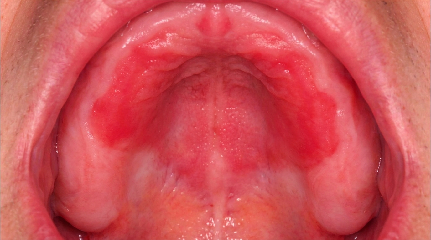
Evaluating the clinical differences between mechanical and fungal mucosal conditions ensures targeted therapy.
Clinical Parameter | Traumatic Denture Sore (Decubitus Ulcer) | Denture Stomatitis (Candida-Induced) |
Primary Etiology | Mechanical over-extension or occlusal high spot | Fungal biofilm (Candida albicans) on acrylic |
Visual Appearance | Localized, deep ulcer with gray center and red rim | Diffuse, bright red velvety erythema under base |
Pain Level | Sharp, stabbing, acute pain upon chewing | Mild burning sensation, dryness, or painless |
Common Location | Vestibular sulcus margins, sharp bone ridges | Palatal vault beneath full maxillary denture |
Clinical Treatment | Selective acrylic relief with burs, soft relining | Topical antifungals and chemical denture soaks |
Healing Duration | Rapid; 48 to 72 hours after pressure relief | 10 to 14 days of antifungal compliance |
Frequently Asked Questions
How do I know if my sore gum from dentures is normal or an infection?
A normal sore gum from dentures is typically localized to one or two distinct tender spots where the acrylic rubs tightly against the gum, appearing as a small ulcer with a white or gray center and a red border. An infection, such as denture stomatitis, manifests as widespread, bright red inflammation covering the entire roof of your mouth, often accompanied by a persistent burning sensation, bad breath, or cracked corners of the lips.
What is the fastest way to heal denture sores without ruining my denture?
The fastest way is to visit your dentist for a minor chairside adjustment. The dentist uses specialized pressure-indicating paste to identify and buff away the exact millimeter of excess plastic causing the friction. Combine this with removing your denture for several hours, rinsing with warm salt water, and applying a protective oral gel; the sore will typically heal within forty-eight to seventy-two hours.
What to use for denture sores to relieve pain while eating?
To relieve pain while eating, apply an over-the-counter topical oral anesthetic gel containing benzocaine or a soothing hyaluronic acid cream directly to the sore spot fifteen minutes before your meal. However, this is only a temporary bridge; if the denture continues digging into the gum, professional acrylic relief is essential.
Can denture stomatitis spread to other parts of my body?
In healthy individuals, denture stomatitis remains localized to the oral mucosa beneath the prosthetic base. However, in immunocompromised patients, elderly individuals, or people with uncontrolled diabetes, untreated oral fungal overgrowth can lead to esophageal candidiasis or secondary angular cheilitis at the corners of the mouth, requiring prompt medical intervention.
Why do I suddenly have a sore from dentures that I have worn for years?
Developing a sore under an old, familiar denture happens because jawbone continuously shrinks and reshapes over time through natural alveolar resorption. As the bone recedes, the rigid denture base no longer matches the tissue contours, causing the appliance to wobble, tilt, and create new friction points. An old denture causing sores usually needs a professional laboratory reline or complete replacement.
About the Author