




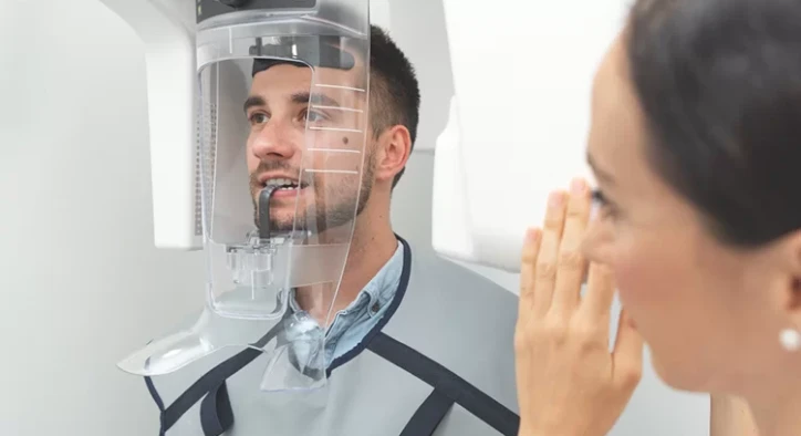

A panoramic X-ray dental image is one of the first things many patients see at a new dental clinic. It is a single wide picture that shows all the teeth, both jaws and the structures around them, and it often takes less time to capture than it takes to fill in the registration form. Dentists use it to get a general map of the mouth before deciding what other tests or treatments are needed.
This guide explains what a panoramic dental X-ray is, how it is taken and what it can and cannot show. It also covers how safe the procedure is, when dentists recommend it before implants, orthodontic work or wisdom tooth removal, how it differs from a three dimensional scan and how to make sure the image is clear enough to be useful.
Table Of Contents
- What Is a Panoramic Dental X-Ray?
- How Is a Panoramic X-Ray Taken?
- What Does a Panoramic Dental X-Ray Show?
- What a Panoramic X-Ray Cannot Show
- When Do Dentists Recommend a Panoramic X-Ray?
- Panoramic X-Ray for Dental Implants
- Panoramic X-Ray in Orthodontics and Wisdom Tooth Removal
- Is a Panoramic Dental X-Ray Safe?
- How to Get a Clear Panoramic X-Ray
- Understanding Your Panoramic X-Ray
- How Often Do You Need a Panoramic X-Ray?
- Frequently Asked Questions
What Is a Panoramic Dental X-Ray?
A panoramic X-ray, also called an OPG or orthopantomogram, is an extraoral image, which means the film or sensor stays outside the mouth. During the exposure, the X-ray source and the detector rotate around the head in a half circle and record a curved slice of the jaws. The result is a flat, wide image that shows the upper and lower teeth, the jawbones, the jaw joints and the sinuses together.
Because everything appears on one picture, the panoramic X-ray is often called the overview image of dentistry. It does not replace close up images of individual teeth, but it gives the dentist a quick and wide look at areas that would otherwise need many separate pictures.
How Is a Panoramic X-Ray Taken?
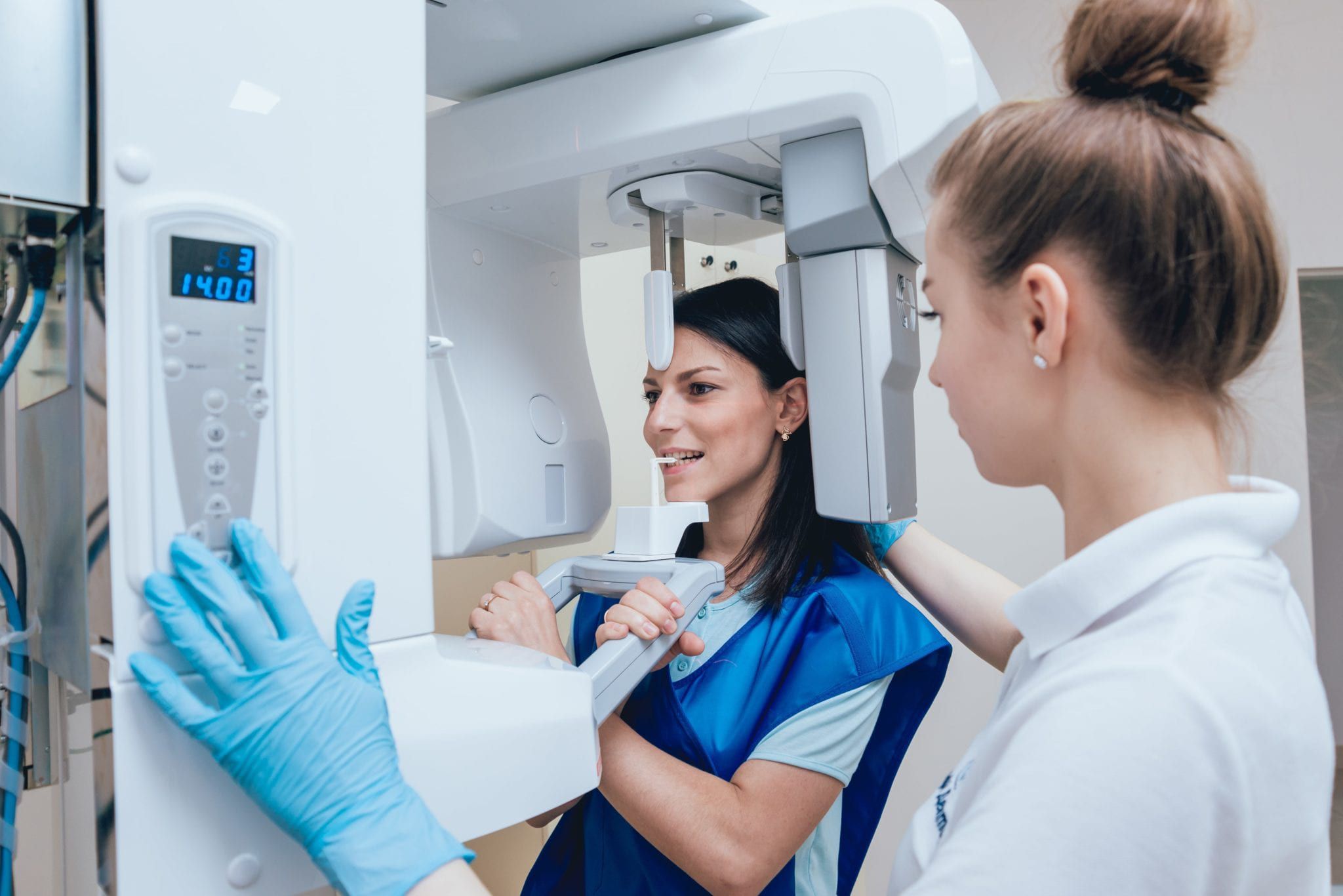
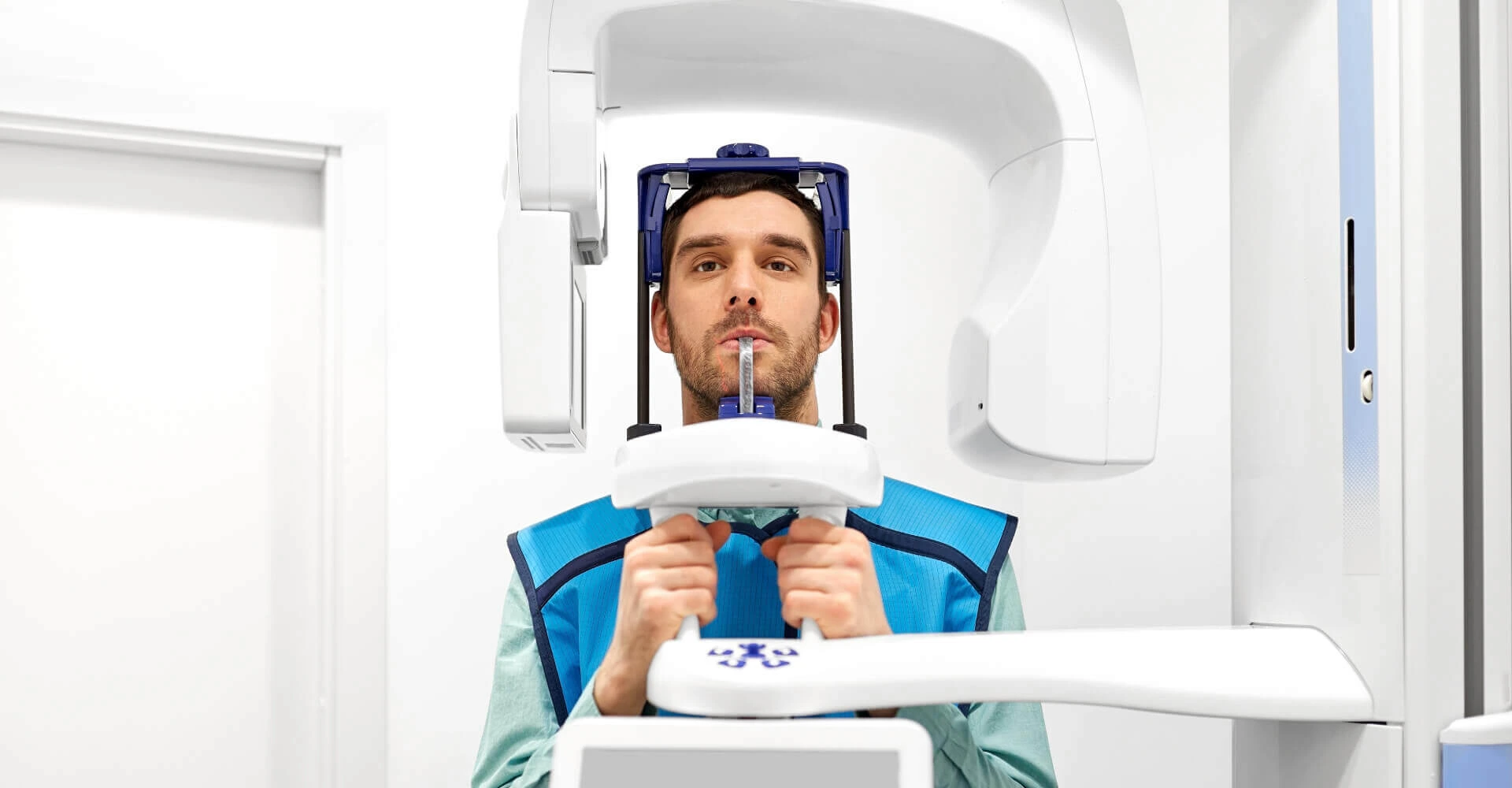
The procedure is simple and painless. You remove earrings, glasses, hairpins and any removable dental appliances, because metal can create shadows on the image. You then stand or sit in the machine, bite lightly on a small plastic guide that keeps the front teeth in position and rest your chin on a support. The technician lines your head up with light guides, and this step matters because a tilted head can distort the image.
The machine moves around your head for about ten to fifteen seconds, and you need to stay completely still during this time. You place your tongue on the roof of your mouth to prevent a dark band of air from covering the upper teeth. The image appears on a screen within seconds, and the dentist checks it immediately to confirm that nothing needs to be repeated.
What Does a Panoramic Dental X-Ray Show?
The image shows the number and position of the teeth, including teeth that have not yet come through and wisdom teeth that are still under the gum. It shows missing teeth, crowns, bridges, implants and fillings, and it gives a general view of the bone height around the teeth. A dentist can recognise large cavities, deep decay, root canal treatments, cysts and signs of bone loss from gum disease.
It also shows structures that are hard to see on small images. The jaw joints can be checked for obvious changes, the sinus floors can be seen in relation to the upper back teeth and the course of the nerve canal in the lower jaw can be traced. This is why the panoramic X-ray is the standard first image before planning wisdom tooth removal, implants or orthodontic treatment.
What a Panoramic X-Ray Cannot Show
A panoramic image is two dimensional, so it compresses the mouth into a flat picture. It does not show the width of the jawbone from cheek to tongue, and bone height can appear larger or smaller than in reality because the image is slightly magnified and can be distorted at the edges. This is the main reason why implant surgery is planned on a three dimensional scan.
Small cavities between neighbouring teeth, early cracks, early gum disease and the condition of the soft tissue are also difficult to judge. Teeth that overlap on the image can hide each other, and the front teeth are often less sharp than on close up pictures. Tooth colour, shape and bite cannot be assessed from an X-ray at all, which is why the image is always read together with a clinical examination.
| Type of image | What it covers | Best used for |
|---|---|---|
| Panoramic X-ray | All teeth, both jaws, joints and sinuses | General overview, wisdom teeth, treatment planning |
| Bitewing X-ray | Crowns of the back teeth, upper and lower together | Cavities between teeth, bone level |
| Periapical X-ray | One or two teeth from crown to root tip | Root canal problems, infections, detail |
| Cephalometric X-ray | Side view of the skull | Orthodontic analysis |
| 3D cone beam scan | A three dimensional volume of the jaws | Implants, nerve position, complex surgery |
When Do Dentists Recommend a Panoramic X-Ray?
Most clinics take a panoramic X-ray at the first visit of a new patient, because it gives a baseline record of the mouth. It is repeated when something changes, for example after trauma, when a new problem is suspected or every few years in a patient with a history of dental disease. For children, it helps to follow the development of the permanent teeth and to see whether there is enough room for them.
It is also requested before specific treatments. Wisdom tooth removal, orthodontic treatment, implant planning, root canal treatment on a back tooth and the assessment of a cyst or an unexplained jaw pain all start with a panoramic image. In each case, the dentist decides whether it is enough or whether a closer or three dimensional image is needed.
Panoramic X-Ray for Dental Implants
For dental implants, the panoramic X-ray is the first step and not the last. It shows the number of missing teeth, the approximate bone height, the position of the sinus in the upper jaw and the nerve canal in the lower jaw. With this information, the dentist can tell whether implants are likely to be possible and which areas need a closer look.
The final plan needs a three dimensional cone beam scan, because the width of the bone and the exact distance to the nerve must be measured. Many clinics therefore use both images, with the panoramic X-ray for orientation and the scan for the exact placement of each implant.
Panoramic X-Ray in Orthodontics and Wisdom Tooth Removal
In orthodontics, the panoramic image shows whether all permanent teeth are present, whether any teeth are missing or extra, and where unerupted teeth such as canines are located. It helps to decide whether teeth need to be removed to create space and how the roots are positioned. A side view of the skull is usually added for measurements of the jaw relationship.
For wisdom teeth, the image shows how deep the teeth lie, in which direction they are tilted and how close the roots are to the nerve in the lower jaw. This helps the dentist or surgeon decide how difficult the removal will be and whether a three dimensional scan is needed. When the roots lie right next to the nerve, an additional scan makes the surgery safer.
Is a Panoramic Dental X-Ray Safe?
A panoramic X-ray uses a low dose of radiation, which is similar to or lower than the natural background radiation a person receives over a few days. The dose is lower than that of a medical CT scan of the head and comparable to a short flight. Modern digital machines have reduced the dose further, and the examination is considered safe when it is done for a good reason.
Dentists follow the principle of using the lowest dose that gives the information needed, and they do not take images without a clinical purpose. A lead apron is often used in addition, and pregnant patients should always tell the dentist, so that non urgent images can be postponed. Children receive a reduced setting that is adapted to the size of the head.
How to Get a Clear Panoramic X-Ray
A clear image depends partly on you. Remove all metal objects from the head and neck area, including piercings and necklaces, and tell the technician about any implants or bridges. Stand upright with the spine straight and keep your head still, as even a small movement blurs the image.
Keep your tongue pressed against the roof of the mouth, bite on the guide in the right position and do not swallow while the machine is turning. If the image is unclear, tilted or cut off, the dentist may ask you to repeat it. A good quality image is worth the extra minute, because planning and diagnosis depend on it.
Understanding Your Panoramic X-Ray
Looking at your own image can be interesting, but it is easy to misread. Teeth and fillings appear white, bone appears grey and air spaces such as the sinuses and the airway appear dark. Healthy bone has a fairly even grey tone, and areas of loss or infection show up as darker patches.
Some normal structures can look like problems to a non specialist, such as the shadows of the tongue, the spine and the jaw joints. A dentist reads the image together with the clinical findings and explains it on the screen. If you send the file to another clinic for a second opinion, always send the original digital file and not a photograph of the screen, because detail is lost in a photo.
How Often Do You Need a Panoramic X-Ray?
There is no fixed interval, and it depends on the patient. Healthy adults with no new problems may need one only every three to five years, while patients in active treatment or with a high risk of dental disease may need it more often. Children are checked during growth at the dentist's discretion.
If you move to a new dentist, bringing a recent image can avoid a repeat exposure. Digital images can be stored and shared easily, and keeping a copy of your own files helps if you receive treatment in more than one place or country.
Frequently Asked Questions
What is a panoramic X-ray used for in dentistry?
It is used to give an overview of all teeth, both jaws, the jaw joints and the sinuses in one image. Dentists use it as a baseline and to plan treatments such as implants, orthodontics and wisdom tooth removal.
Is a panoramic dental X-ray painful or dangerous?
No. The procedure is painless and takes only seconds. The radiation dose is low, and the examination is considered safe when it is done for a clear clinical reason.
Can a panoramic X-ray detect cavities?
It can show large cavities and deep decay, but small cavities between teeth are often missed. Bitewing and close up X-rays are more reliable for this purpose.
Is a panoramic X-ray enough for dental implants?
It is enough for a first assessment of whether implants may be possible. The final plan needs a three dimensional scan to measure the width of the bone and the position of the nerve.
Can I take a panoramic X-ray during pregnancy?
Routine images are usually postponed, and urgent imaging is done with protective measures when the benefit is greater than the risk. Always tell the dentist if you are pregnant or think you might be.
O autorovi