




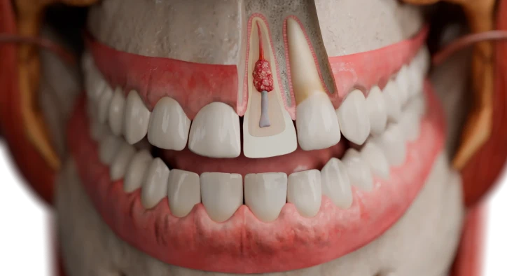

The progressive loss of mineralized dental root structure caused by specialized clastic cell activity represents one of the most challenging conditions in endodontics and dental traumatology. When evaluating progressive root shortening or internal pulp wall expansion on radiographic imaging, answering what is tooth root resorption is the fundamental first step toward saving the natural dentition. While physiological resorption is a healthy developmental event in children, pathological destruction in permanent teeth can compromise structural anchoring and lead to early tooth loss if left untreated.
For patients experiencing localized tooth mobility, unexplained pink spots on the crown, or subtle throbbing discomfort, identifying tooth root resorption symptoms early is critical for successful clinical intervention. Because the early stages of clastic destruction are often completely asymptomatic, routine high-resolution 3D imaging plays a crucial role in preventing structural collapse. This comprehensive clinical guide explores the cellular mechanisms of root breakdown, examines what causes tooth root resorption, differentiates between physiological primary tooth root resorption and pathological tooth root resorption in adults, assesses the risks associated with wisdom tooth root resorption, and outlines modern protocols for tooth root resorption treatment.
Table Of Contents
- What Is Tooth Root Resorption and How Does It Occur?
- What Causes Tooth Root Resorption and How Do Types Differ?
- What Are the Typical Tooth Root Resorption Symptoms?
- Restorative & Endodontic Solutions: Tooth Root Resorption Treatment
- Structural Evaluation: Normal Root vs Resorbed Root State
- Frequently Asked Questions
What Is Tooth Root Resorption and How Does It Occur?
To establish a proper clinical diagnosis, dentists and endodontists evaluate the cellular mechanisms driving tissue breakdown. When explaining what is tooth root resorption, specialists describe a biological process where specialized multinucleated cells called odontoclasts dissolve the mineralized cementum and dentin of the root surface.
The biological progression follows two distinct pathways:
- [ PROTECTIVE LAYER DAMAGE ]: Physical trauma or chronic infection compromises cementum/predentin
- [ ODONTOCLAST ACTIVATION ]: Multinucleated clastic cells recruit to the denuded root surface
- [ MINERAL DISSOLUTION ]: Acid and proteolytic enzymes actively dissolve cementum and dentin
- [ RESORPTION CAVITY ]: Root structure shortens externally or expands internally into the pulp
Under healthy conditions, the root surface is protected externally by the cementum and periodontal ligament, and internally by the unmineralized predentin layer. When physical injury, excessive pressure, or chronic inflammation disrupts these protective barriers, odontoclasts attach to the exposed mineralized structure, triggering active tissue breakdown.
What Causes Tooth Root Resorption and How Do Types Differ?
Understanding what causes tooth root resorption requires distinguishing between physiological processes in primary teeth and pathological destructive conditions in adult dentition.
The primary categories include:
- Primary Tooth Root Resorption (Physiological): In growing children, primary tooth root resorption is a natural and necessary biological process. As the permanent tooth erupts beneath the baby tooth, pressure activates odontoclasts to resorb the primary roots, allowing the baby tooth to loosen and shed naturally.
- Pathological Tooth Root Resorption in Adults: In permanent teeth, root loss is always pathological. It is primarily triggered by dental trauma (such as sports injuries or falls), excessive and rapid orthodontic forces during braces treatment, chronic intracanal pulp infections, or pressure from adjacent impacted teeth.
- Wisdom Tooth Root Resorption: Horizontally or mesioangularly impacted wisdom teeth frequently exert heavy physical pressure against the roots of the adjacent second molars, causing severe wisdom tooth root resorption that often necessitates extracting the wisdom tooth to save the molar.
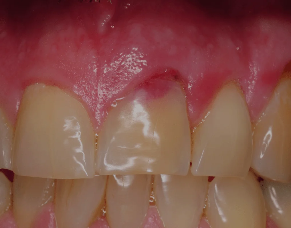
- Internal vs. External Resorption: Internal resorption begins inside the pulp chamber and hollows out the tooth from the inside out, often creating a characteristic pink spot on the enamel. External resorption originates on the outer root surface from the periodontal ligament and eats inward toward the pulp.
What Are the Typical Tooth Root Resorption Symptoms?
In its early stages, pathological root resorption is almost always painless and silent, making regular dental examinations and radiographic checks essential.
As the lesion advances, characteristic tooth root resorption symptoms begin to emerge:
- Pink Tooth Appearance: In cases of internal cervical resorption, highly vascularized granulation tissue inside the pulp chamber shines through the thinned enamel, creating a distinctive pink hue.
- Unexplained Tooth Mobility: As the root structure progressively shortens and loses its periodontal ligament attachment, the tooth becomes loose.
- Dull Aching Pain and Pressure: If external resorption triggers secondary periodontal inflammation or if internal resorption breaches the pulp, patients experience persistent throbbing pain and temperature sensitivity.
- Swelling along the Gumline: Localized soft-tissue swelling or sinus tracts can develop adjacent to the resorptive defect.
Restorative & Endodontic Solutions: Tooth Root Resorption Treatment
Selecting the appropriate tooth root resorption treatment depends on whether the defect is located internally within the canal or externally along the outer root surface.
- [ INTERNAL RESORPTION ]: Complete root canal therapy + Bioceramic / MTA obturation
- [ EXTERNAL CERVICAL ]: Surgical flap reflection + Defect debridement + Composite/Biodentine seal
- [ SEVERE APICAL LOSS ]: Occlusal force reduction + Splinting or extraction and dental implant
Modern clinical protocols include:
- Advanced Bioceramic Root Canal Therapy: For internal resorption, the endodontist removes the hyperactive pulp tissue, disinfects the complex irregular canal space with sonic irrigation, and fills the hollowed cavity with Mineral Trioxide Aggregate (MTA) or bioceramic materials to halt destruction.
- Micro-Surgical Defect Repair: For external cervical resorption, a periodontist gently reflects a gum flap, cleans out the destructive clastic tissue from the root surface, and seals the defect with resin-modified glass ionomer or Biodentine before repositioning the gum.
- Extraction of Impacted Teeth: In cases of wisdom tooth root resorption, the offending impacted third molar is surgically removed immediately to eliminate pressure and arrest further damage to the adjacent tooth.
- Prosthetic Replacement: When tooth root resorption in adults has destroyed more than 50% of the root structure, the tooth is structurally unsalvageable and is replaced with a dental implant.
Structural Evaluation: Normal Root vs Resorbed Root State
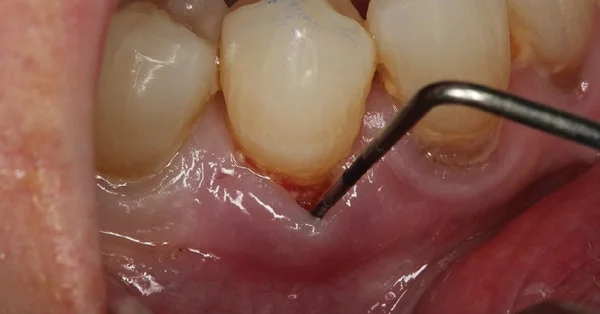
Analyzing the clinical differences between healthy root anatomy and progressive resorptive states highlights the importance of early diagnosis.
Clinical Parameter | Healthy Intact Root Architecture | Resorbed Root Structure |
Root Length and Volume | Full anatomical length; robust bone anchoring | Shortened, blunted, or hollowed root structure |
Pulp Wall Integrity | Smooth, continuous canal borders | Irregular ballooning or invasive exterior defects |
Periodontal Ligament Space | Uniform, thin radiolucent line along root | Disrupted ligament space; localized bone replacement |
Tooth Stability Index | Firmly anchored; zero pathological mobility | Increased mobility; high risk of structural fracture |
Frequently Asked Questions
Can tooth root resorption be stopped once it starts?
Yes. If detected early through routine dental radiographs or 3D CBCT scans, root resorption can be completely arrested. For internal resorption, root canal therapy with bioceramic sealing stops clastic activity immediately. For external resorption, surgical debridement and eliminating causative factors (such as excessive orthodontic pressure or impacted wisdom teeth) halts progression.
Will a tooth with root resorption eventually fall out?
If left untreated, progressive external or internal resorption will eventually destroy the root anchor, resulting in severe mobility, bacterial infection, structural fracture, and tooth loss. However, prompt treatment preserves the tooth for decades.
Why do braces sometimes cause tooth root resorption in adults?
Orthodontic movement relies on controlled mechanical pressure to remodel bone. If orthodontic forces are applied too rapidly or excessively, or if a patient has a genetic predisposition, the pressure can compress the periodontal ligament, damaging the protective cementum and triggering localized apical root shortening.
Is tooth root resorption treatment painful?
No. Treatment is performed under local anesthesia, ensuring you experience no pain during the procedure. Root canal therapy and minor microsurgical repairs are performed with high-precision surgical microscopes, resulting in minimal post-operative discomfort that is easily managed with standard pain relievers.
How is root resorption diagnosed if there are no symptoms?
Because root resorption is typically painless in its early stages, it is most often diagnosed during routine bitewing or panoramic dental X-rays. For precise 3D evaluation, clinicians use Cone-Beam Computed Tomography (CBCT) to determine the exact location, depth, and structural extent of the resorptive defect.
About the Author