




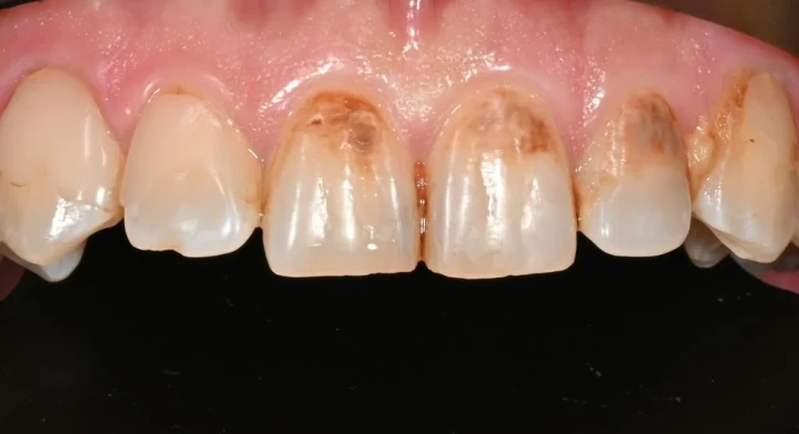

The structural integrity of dental enamel represents the body's first line of defense against mechanical masticatory wear, bacterial acids, and temperature fluctuations. When genetic anomalies, prenatal disruptions, or early childhood illnesses interfere with enamel formation, understanding what is enamel hypoplasia provides the clinical foundation for preserving damaged natural crowns. Unlike dental erosion or acid demineralization, dental enamel hypoplasia is a developmental disturbance that occurs while teeth are still forming beneath the gums, resulting in teeth that erupt with thin, grooved, pitted, or missing enamel layers.
For parents observing chalky white spots or yellow pits in their child's mouth, evaluating enamel hypoplasia baby teeth early is critical for preventing rampant childhood decay. Similarly, adult patients frequently ask does enamel hypoplasia affect adult teeth, research complex systemic enamel hypoplasia causes, and seek effective options for enamel hypoplasia treatment. By exploring documented transformations in enamel hypoplasia before and after cases, clinicians can implement minimally invasive resin therapies or high-strength ceramic restorations to restore natural aesthetics and structural durability. This comprehensive clinical guide explores ameloblast cellular biology, systemic risk factors, preventative strategies, and modern restorative protocols.
Table Of Contents
- What Is Enamel Hypoplasia and How Does It Develop?
- Etiological Factors: What Causes Enamel Hypoplasia?
- Clinical Scope: Does Enamel Hypoplasia Affect Adult Teeth and Baby Teeth?
- Structural Evaluation: Enamel Hypoplasia Before and After Restorative Treatment
- Restorative Strategies: Modern Enamel Hypoplasia Treatment Protocols
- Frequently Asked Questions
What Is Enamel Hypoplasia and How Does It Develop?
To understand how developmental enamel defects originate, answering what is enamel hypoplasia requires analyzing the cellular lifecycle of ameloblasts (the specialized cells responsible for producing enamel matrix). Dental enamel hypoplasia is a quantitative defect characterized by an insufficient quantity of enamel formed during the secretory phase of amelogenesis, leaving the underlying dentin poorly shielded.
The cellular and biological breakdown follows a defined pathway:
- [ SYSTEMIC INSULT / GENETIC MUTATION ]: Prenatal, perinatal, or childhood disruption occurs
- [ AMELOBLAST SECRETORY FAILURE ]: Enamel-forming cells become damaged or undergo apoptosis
- [ DEFICIENT ENAMEL MATRIX ]: Inadequate thickness of organic matrix scaffold laid down
- [ INCOMPLETE MINERALIZATION ]: Mineral crystals fail to reach full biological thickness
- [ ERUPTION OF DEFECTIVE CROWN ]: Tooth emerges with visible pits, grooves, or missing enamel
Unlike bones or dentin, dental enamel contains no living cells once fully formed and cannot regenerate, repair, or replace itself biologically. When ameloblasts are injured during early tooth development, the physical defect in the enamel matrix becomes permanently locked into the tooth structure upon eruption.
Etiological Factors: What Causes Enamel Hypoplasia?

Understanding what causes enamel hypoplasia requires distinguishing between localized environmental trauma and broad systemic or hereditary conditions.
The primary clinical triggers include:
- Prenatal and Perinatal Complications: Maternal nutritional deficiencies (especially vitamin D, calcium, and vitamin A), gestational diabetes, maternal smoking, premature birth, low birth weight, and birth hypoxia severely disrupt early ameloblast activity.
- Childhood Systemic Illnesses: Severe high fevers during the first six years of life (measles, chickenpox, scarlet fever), untreated celiac disease causing malabsorption, severe neonatal jaundice, and chronic respiratory infections.
- Localized Physical Trauma (Turner's Hypoplasia): Physical trauma to a primary baby tooth can push its root tip directly into the developing permanent tooth germ beneath, causing localized hypoplasia on that specific adult tooth.
- Genetic Conditions: Inherited conditions like Amelogenesis Imperfecta disrupt enamel matrix genes, causing widespread structural defects across the entire dentition.
- Excessive Fluoride Intake (Severe Fluorosis): Extremely high systemic fluoride ingestion during tooth development can disrupt ameloblast function, creating hypomineralized, pitted enamel zones.
Clinical Scope: Does Enamel Hypoplasia Affect Adult Teeth and Baby Teeth?
Patients and parents frequently ask whether this developmental defect is confined to childhood or does enamel hypoplasia affect adult teeth. Clinical evidence demonstrates that hypoplasia can compromise both dental phases depending on the exact timing of the biological insult.
- [ IN UTERO TO 1 YEAR OF AGE ]: Disruption impacts primary baby teeth and permanent first molars
- [ 1 YEAR TO 6 YEARS OF AGE ]: Disruption impacts permanent incisors, canines, and premolars
When evaluating enamel hypoplasia baby teeth, defects usually manifest as thin, chalky, or yellowish enamel on primary incisors and first primary molars. Because primary teeth naturally possess thinner enamel than permanent teeth, hypoplastic baby teeth break down rapidly under bacterial attacks, leading to early childhood caries.
In contrast, hypoplasia in adult teeth develops when environmental or systemic disruptions occur between the ages of one and six years. Adult teeth emerge with localized deep horizontal grooves, yellow-brown rough patches, or severe incisal pitting that causes acute cold sensitivity and rapid attrition.
Structural Evaluation: Enamel Hypoplasia Before and After Restorative Treatment
Analyzing clinical documentation in enamel hypoplasia before and after cases demonstrates how tailored restorative protocols restore strength, seal sensitive dentin, and rebuild natural smile aesthetics.
Clinical Parameter | Pre-Treatment Hypoplastic State | Post-Treatment Restored State |
Enamel Thickness & Texture | Thin, pitted, grooved, or chipped surface | Smooth, continuous, anatomically contoured surface |
Dentin Sensitivity & Pain | Sharp pain with cold, hot, or acidic food | Zero sensitivity; open dentinal tubules sealed |
Caries Vulnerability | High; porous enamel traps plaque easily | Minimized; non-porous restorative materials |
Aesthetic Integration | Unsightly yellow, brown, or chalky white spots | Natural, lifelike shade matching adjacent enamel |
Restorative Strategies: Modern Enamel Hypoplasia Treatment Protocols

Selecting the most effective enamel hypoplasia treatment depends on the patient's age, the depth of the defect, and the severity of mechanical wear:
- Topical Remineralization & Fluoride Therapy: For mild hypoplasia presenting as superficial white spots without structural enamel loss, regular applications of high-concentration fluoride varnishes, casein phosphopeptide-amorphous calcium phosphate (CPP-ACP), and nano-hydroxyapatite pastes help harden the defective enamel.
- Resin Infiltration (Icon Technique): A micro-invasive procedure where mild hydrochloric acid erodes the porous surface layer, allowing a highly fluid light-cured resin to seep deep into the porous enamel matrix. This blocks stain penetration, stops early decay, and restores a natural, uniform tooth color without drilling.
- Micro-Hybrid Direct Composite Bonding: For localized pits, grooves, or chipped edges, composite resin is applied directly over the conditioned enamel to rebuild normal tooth anatomy and seal exposed dentin.
- Pediatric Stainless Steel or Zirconia Crowns: For severely hypoplastic primary baby molars prone to structural collapse, preformed pediatric crowns protect the tooth until natural shedding occurs.
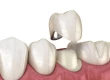
- Porcelain Veneers and Full-Coverage Ceramic Crowns: In adult patients with extensive hypoplasia across the smile line, custom micro-layered lithium disilicate (E-Max) veneers or monolithic zirconia crowns provide permanent aesthetic transformation and maximum fracture resistance.
Frequently Asked Questions
Can enamel hypoplasia be cured or reversed naturally?
No. Once a tooth erupts with defective or missing enamel, the lost mineralized tissue cannot grow back or regenerate naturally because ameloblasts die before the tooth emerges into the mouth. However, modern restorative treatments like resin infiltration, composite bonding, and ceramic restorations can permanently replace lost enamel, seal the tooth, and restore normal function.
Is enamel hypoplasia the same as tooth decay?
No. Enamel hypoplasia is a developmental defect that occurs before the tooth erupts, resulting in a thin or incomplete enamel layer. Tooth decay (caries) is an infectious bacterial disease that destroys healthy enamel after the tooth has erupted. However, teeth with enamel hypoplasia are far more vulnerable to developing rapid tooth decay because their protective enamel barrier is structurally weak.
How can parents prevent enamel hypoplasia in children?
Prevention focuses on early maternal and childhood health: ensuring adequate maternal nutrition with vitamin D, calcium, and iron during pregnancy; avoiding unnecessary medications during gestation; seeking prompt pediatric treatment for high fevers and systemic infections; monitoring systemic fluoride intake; and scheduling a child's first dental visit by age one to identify developmental anomalies early.
Does enamel hypoplasia cause severe tooth pain?
Yes, it can. Because hypoplastic enamel is thin, pitted, or missing in localized areas, the underlying dentin and microscopic dentinal tubules are often left exposed to the oral environment. This direct pathway to the dental nerve can cause acute sensitivity or sharp pain when consuming cold, hot, sweet, or acidic foods.
Why are baby teeth with enamel hypoplasia restored rather than extracted?
Preserving primary baby teeth is critical for proper speech development, comfortable chewing, and maintaining space in the jaw for permanent adult teeth to erupt properly. Extracting hypoplastic baby teeth prematurely causes adjacent teeth to drift, resulting in severe orthodontic crowding, impactions, and bite collapse in the permanent dentition.
Über den Autor