





Discovering that a tooth has lost its normal vitality or seeing a dentist examine a dental radiograph with visible concern often leads to unexpected findings. In modern restorative dentistry, a calcified tooth represents an altered biological state where the inner pulp chamber and root canal spaces become constricted or fully obliterated by dense hard tissue. Rather than an acute bacterial infection, this calcific metamorphosis is typically a defensive biological response to physical trauma or chronic long-term irritation, during which specialized dental cells produce mineralized layers inside the tooth.
Patients presented with this diagnosis frequently ask what is a calcified tooth and seek clear information regarding calcified tooth root causes to understand why their root space narrowed. Many examine calcified tooth symptoms when noticing a single tooth turning yellow or opaque, while others worry about developing a calcified tooth root abscess beneath a blocked root. When confronted with complex treatment plans, patients routinely ask does calcified tooth need root canal therapy, wonder how changes to the calcified tooth layer and internal calcified tooth pulp affect nerve function, and ask with urgency can a calcified tooth be saved from extraction. This clinical guide delivers transparent medical facts to navigate your treatment safely.
Table Of Contents
- Biological Architecture: What Is a Calcified Tooth and How Does Obliteration Form?
- Etiology: Identifying Primary Calcified Tooth Root Causes
- Symptom Profile: Recognizing Distinct Calcified Tooth Symptoms
- Clinical Complications: Can a Calcified Tooth Root Abscess Develop?
- Treatment Decisions: Does a Calcified Tooth Need Root Canal Therapy?
- Endodontic Techniques: Can a Calcified Tooth Be Saved?
- Comprehensive Comparison: Healthy Pulp vs. Calcified Pulp vs. Necrotic Pulp
- Frequently Asked Questions
- What is a calcified tooth and why does it look yellow?
- Does a calcified tooth need root canal therapy if it does not hurt?
- Can a calcified tooth be saved if the canal is completely blocked?
- What are the primary calcified tooth symptoms I should watch for?
- How does a dentist treat a calcified tooth root abscess?
Biological Architecture: What Is a Calcified Tooth and How Does Obliteration Form?
To understand the biological mechanism, one must ask: what is a calcified tooth? In a healthy tooth, the outer enamel and dentin shell encase a hollow interior filled with living, vascularized tissue known as the dental pulp. The pulp contains sensory nerves, blood vessels, and specialized cells called odontoblasts that line the inner dentin wall.
- [ DENTAL TRAUMA OR CHRONIC STRESS ]: Impact, deep restoration, or gradual aging
- [ PULP CANAL OBLITERATION ]: Mineralized dentin layers constrict the pulp space
When a tooth experiences a traumatic concussive blow, a deep restoration, or advanced physiological aging, the neurovascular bundle at the root apex is stressed. In response, the body attempts to protect the nerve by laying down irregular secondary and tertiary dentin along the walls of the pulp chamber. Over months or years, this mineral deposition thickens the inner calcified tooth layer, gradually squeezing the calcified tooth pulp until the root canal appears completely invisible on standard two-dimensional radiographs. This condition is clinically recognized as pulp canal obliteration.
Identifying Primary Calcified Tooth Root Causes
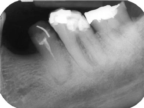
Analyzing the specific calcified tooth root causes allows dental clinicians to predict the structural behavior of the root canal system and formulate appropriate monitoring plans.
- Physical Dental Trauma: A blunt impact, such as a sports collision, bicycle fall, or direct blow to the mouth, is the leading trigger, especially in anterior incisors during adolescence. Even if the tooth does not crack or dislodge, the physical shock disrupts pulpal blood supply, stimulating rapid reparative dentin formation.
- Physiological Aging: Throughout human life, odontoblasts deposit secondary dentin at a slow, continuous rate. In elderly individuals, it is clinically normal for the pulp chamber to shrink significantly, narrowing the root canals throughout the entire mouth.
- Extensive Restorative History: Teeth that have undergone multiple deep fillings, crown preparations, or prolonged orthodontic tooth movement experience chronic, low-grade physical stress, encouraging reactionary calcification.
- Pulp Stones (Denticles): Discrete, localized calcified nodules can form spontaneously inside the coronal pulp chamber in response to local genetic tendencies or systemic metabolic factors, complicating canal entrance visibility.
Symptom Profile: Recognizing Distinct Calcified Tooth Symptoms
Understanding calcified tooth symptoms is essential because calcific metamorphosis behaves differently from ordinary bacterial tooth decay.
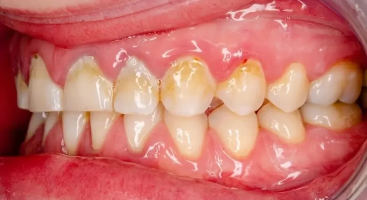
[ CORONAL OPACITY & YELLOWING ]: Thickened dentin blocks light, creating a darker, amber hue
[ SILENT PULPAL STABILITY ]: Tooth remains asymptomatic without pain in 80 to 90 percent of cases
- Amber or Yellow Discoloration: The most prominent visual sign. As the pulp chamber fills with dense, opaque tertiary dentin, light no longer transmits through the crown in the same manner, making the tooth appear distinctly darker, yellow, or grayish-amber compared to adjacent teeth.
- Loss of Cold and Electrical Sensitivity: Because mineral deposits insulate the sensory nerves, the tooth typically shows a delayed response or complete lack of sensation during electric pulp testing and cold thermal tests, mimicking a non-vital tooth.
- Absence of Acute Pain: In eighty to ninety percent of cases, a calcified tooth remains completely comfortable, functioning naturally during mastication without throbbing, lingering aches, or chewing pain.
- Delayed Periapical Tenderness: If the residual microscopic pulp tissue eventually undergoes ischemic necrosis, bacteria can colonize the apical region, producing tenderness upon tapping or spontaneous dull aching.
Clinical Complications: Can a Calcified Tooth Root Abscess Develop?
A central concern among patients is whether an obliterated tooth can harbor serious infections, specifically developing a calcified tooth root abscess.
While the canal appears solidly filled with bone-like material on radiographs, histology proves that microscopic ribbons of organic soft tissue and tiny canal tracks almost always persist throughout the calcified tooth root. If bacteria from micro-leaking fillings, gum disease, or bloodstream travel enter these microscopic dead-end channels, the body cannot circulate defense cells through the constricted space to fight the microbes. Over time, bacterial toxins accumulate at the apex, producing an apical periodontitis lesion or a painful calcified tooth root abscess accompanied by localized gum swelling, throbbing pressure, or a draining fistula.
Treatment Decisions: Does a Calcified Tooth Need Root Canal Therapy?
When evaluating clinical management, the decisive diagnostic question is: does calcified tooth need root canal therapy automatically upon discovery?
From an evidence-based endodontic standpoint, a calcified tooth does not require a root canal simply because the canal appears obliterated or yellow on an X-ray. If the tooth is completely asymptomatic, exhibits no tenderness to percussion, and shows a pristine, normal periodontal ligament space with zero bone loss around the root apex, the standard of care is conservative observation and yearly radiographic monitoring. Prophylactic root canal treatment on a symptom-free calcified tooth carries high procedural risks, including mechanical root perforation or instrument breakage during drilling.
However, endodontic therapy becomes mandatory under two specific circumstances:
- Presence of Apical Pathology: If radiographs show an osteolytic lesion, dark halo, or bone loss around the apex, or if the patient presents with a calcified tooth root abscess.
- Clinical Symptoms of Pulpitis: If the tooth begins throbbing spontaneously, hurts upon biting down, or exhibits localized mucosal swelling.
Endodontic Techniques: Can a Calcified Tooth Be Saved?
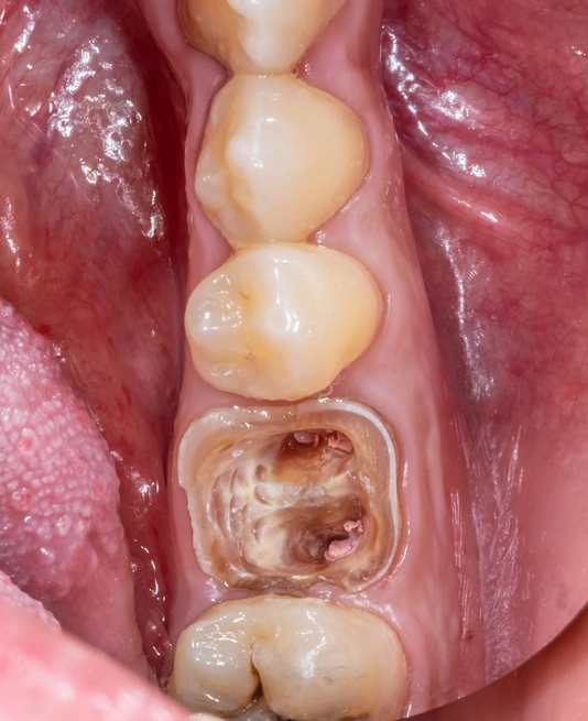
Patients facing complex endodontic treatments routinely ask with great anxiety: can a calcified tooth be saved? The definitive answer is yes; modern endodontic technology allows dental specialists to locate, navigate, and clean seemingly invisible canals with exceptional success rates.
[ HIGH-RESOLUTION 3D CBCT ]: Micron-level mapping of microscopic patent canal paths
[ GUIDED ENDODONTIC NAVIGATION ]: 3D printed drill guide delivers precise, conservative access
- Dental Operating Microscopes: Endodontists utilize high-power surgical microscopes providing up to twenty-five times magnification and coaxial LED illumination. This visual clarity allows clinicians to distinguish between the lighter, yellowish secondary dentin and the darker, natural canal path.
- Ultrasonic Troughing Tips: Rather than using high-speed rotary burs that remove sound dentin aggressively, specialists employ delicate ultrasonic diamond tips to gently shave away calcified obstructions without creating root perforations.
- Chemical Chelating Agents: Liquid solutions of ethylenediaminetetraacetic acid (EDTA) are introduced into the access cavity. EDTA binds to calcium ions, demineralizing and softening the calcified tooth layer to allow micro-instruments to slide into tight spaces.
- Guided 3D Endodontic Templates: In completely obliterated anterior teeth, virtual planning software combines 3D CBCT data with surface scans to print a custom surgical guide. A tiny micro-bur passes through the guide sleeve, drilling a straight, micron-precise path directly into the microscopic canal without removing healthy enamel.
- Aesthetic Internal Bleaching: If the primary complaint is unsightly yellow discoloration on a healthy tooth, non-invasive home bleaching or targeted internal walking bleach techniques restore natural shade matching without requiring aggressive porcelain crown preparations.
Comprehensive Comparison: Healthy Pulp vs. Calcified Pulp vs. Necrotic Pulp
Evaluating the physiological characteristics across different states of the dental pulp clarifies necessary clinical interventions.
Clinical Parameter | Healthy Normal Pulp | Calcified Tooth Pulp (PCO) | Necrotic Pulpal Infection |
Pulp Chamber Space | Wide, patent chamber and canals | Severely constricted or invisible | Patent or partially calcified |
Response to Cold Test | Immediate, transient sensation | Delayed or zero cold sensation | Zero response to cold stimulation |
Tooth Color Tone | Natural translucent white | Yellowish, amber, or opaque | Gray, dark brown, or bluish |
Apical Bone Condition | Intact, healthy bone architecture | Typically normal bone outline | Dark shadow, apical radiolucency |
Spontaneous Pain | None | Absent in most cases | Severe throbbing or pressure |
Treatment Required | Routine preventive maintenance | Periodic observation unless infected | Mandatory root canal or apicoectomy |
Frequently Asked Questions
What is a calcified tooth and why does it look yellow?
A calcified tooth is a tooth where the inner pulp chamber and root canal have become filled with dense reparative dentin layers in response to trauma, deep fillings, or aging. The tooth looks yellow or darker because the thickened, opaque dentin layer inside the crown blocks natural light transmission, reducing the tooth's translucency.
Does a calcified tooth need root canal therapy if it does not hurt?
No. If a calcified tooth has no pain, no swelling, and shows normal healthy bone around the root tip on digital X-rays, it does not need a root canal. Routine clinical monitoring and periodic radiographs are the recommended approach to avoid unnecessary procedural complications.
Can a calcified tooth be saved if the canal is completely blocked?
Yes. Modern endodontists successfully save calcified teeth using surgical operating microscopes, 3D CBCT scans, ultrasonic instruments, and 3D printed guided endodontic templates. These advanced tools allow specialists to locate and negotiate microscopic canal paths that cannot be seen with the naked eye.
What are the primary calcified tooth symptoms I should watch for?
The most common symptoms include noticeable yellowing or darkening of the clinical crown, lack of sensitivity when drinking ice-cold liquids, and a normal appearance on the gum tissue. If an infection develops, you may experience tenderness when chewing, throbbing pressure, or a gum boil near the root tip.
How does a dentist treat a calcified tooth root abscess?
To treat an abscess in a calcified tooth, an endodontist performs a microscopic root canal treatment using 3D imaging and ultrasonic instruments to locate and disinfect the hidden canal pathways. If the canal is impassable through the crown, the specialist performs an apicoectomy, which is a minor surgical procedure to clean the root tip directly through the gum and place a biological seal.
About the Author