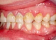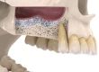





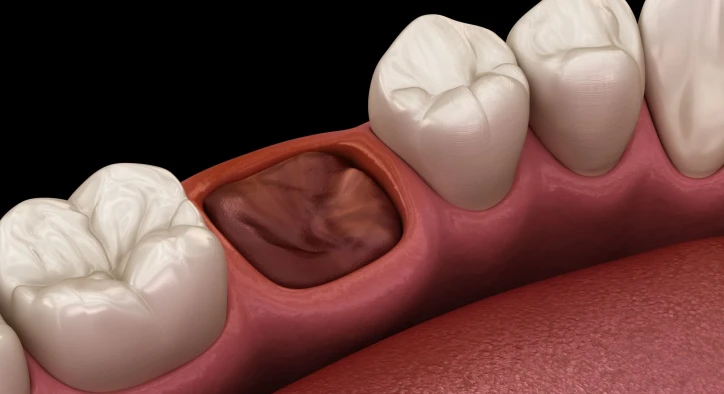

Undergoing a surgical or simple exodontia procedure leaves an open bony wound that requires immediate biological sealing to safeguard underlying tissues. In oral and maxillofacial surgery, the development of a stable tooth extraction clot represents the initial, non-negotiable cornerstone of alveolar bone and soft-tissue regeneration. Rather than serving merely as a temporary plug to arrest bleeding, this initial blood mass acts as a bioactive matrix packed with platelets, cytokines, and cellular signaling factors that drive microvascular reconstruction and osteogenesis inside the empty socket.
Patients closely inspecting their surgical site in a mirror frequently have questions about normal tooth extraction clot formation and wonder how an anterior site differs from a posterior wisdom tooth extraction clot. A common source of alarm occurs when patients observe their tooth extraction clot turning white, mistaking sterile fibrin deposits for purulent infection. Others experience anxiety asking why won't my tooth extraction clot during the first post-operative hours, observe the physical changes of a tooth extraction clot shrinking, or seek confirmation on what a healthy healing tooth extraction clot looks like. This clinical guide delivers transparent medical facts to ensure a safe, predictable recovery.
Table Of Contents
- Biological Foundations: What Is a Tooth Extraction Clot and How Does It Form?
- Healing Stages: Color Shifts, Clot Shrinkage, and White Tissue Formation
- Wisdom Teeth Dynamics: The Complexity of a Wisdom Tooth Extraction Clot
- Etiology: Why Won't My Tooth Extraction Clot Form or Stay Intact?
- Complications: Loss of Clot and Dry Socket Prevention
- Comprehensive Comparison: Healing Clot vs. Dry Socket vs. Socket Infection
- Frequently Asked Questions
- Why is my tooth extraction clot turning white a few days after surgery?
- How long does it take for a tooth extraction clot to become secure?
- What should I do if my extraction socket won't stop bleeding on the first day?
- Why is my tooth extraction clot shrinking and creating a dent in my gum?
- Can drinking through a straw really dislodge a wisdom tooth extraction clot?
Biological Foundations: What Is a Tooth Extraction Clot and How Does It Form?
To understand proper healing, one must examine the hematological events following tooth removal. When the periodontal ligament is severed and the tooth is extracted from the alveolar bone, severed vessels in the surrounding bone marrow flood the socket with blood.
- [ MECHANICAL TOOTH EXTRACTION ]: Severing of periodontal ligament & microvascular network
- [ STABLE ERYTHROCYTE PLUG ]: Biologically seals cortical bone and prevents bacterial invasion
The process of tooth extraction clot formation initiates within seconds. Platelets adhere to exposed collagen fibers along the bony socket walls, releasing adenosine diphosphate and thromboxane to recruit circulating platelets. Simultaneously, the extrinsic and intrinsic coagulation cascades generate thrombin, an enzyme that converts soluble fibrinogen into an insoluble mesh of sticky fibrin strands. These strands trap red blood cells and leukocytes, producing a gelatinous, dark red plug within fifteen to thirty minutes. This biological shield seals exposed bone and insulates sensory nerve endings from air, food debris, and oral bacteria.
Healing Stages: Color Shifts, Clot Shrinkage, and White Tissue Formation
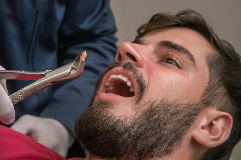
Evaluating the visual transitions of a healing tooth extraction clot over the first two weeks helps patients distinguish healthy cellular turnover from pathological breakdown.
[ DAY 1: FRESH GELATINOUS CLOT ]: Dark red or maroon sponge-like plug filling socket volume
[ DAYS 3 TO 5: FIBRIN MATURATION ]: Whitish, cream-colored, or yellowish non-purulent matrix
[ DAYS 7 TO 14: EPITHELIALIZATION ]: Pink soft tissue contracts; clot shrinks into healthy mucosa
- Day One (Fresh Hemostatic Clot): A deep dark red, gelatinous plug fills the socket. It is fragile and easily dislodged by suction or pressure.
- Days Three to Five (The Fibrin Phase): Patients frequently panic upon noticing their tooth extraction clot turning white or developing a yellowish, creamy surface film. This white layer is not pus, infection, or trapped food. It consists of dead leukocytes and avascular fibrin mesh that replaces superficial red blood cells as granulation tissue ingrowth begins from the bony walls.
- Days Five to Seven (Clot Contraction): The phenomenon of a tooth extraction clot shrinking becomes visibly apparent. Fibroblasts enter the scaffold and synthesize collagen fibers, while myofibroblasts contract the wound perimeter, drawing the gum margins closer together.
- Days Seven to Fourteen (Epithelial Coverage): The clot is almost entirely replaced by highly vascularized granulation tissue, which transitions into immature woven bone underneath while pink surface mucosa seals the crest completely.
Wisdom Teeth Dynamics: The Complexity of a Wisdom Tooth Extraction Clot
Managing a wisdom tooth extraction clot in the posterior mandible presents distinct anatomical challenges compared to anterior extractions. Lower third molars sit within dense cortical bone with lower blood supply than the richly vascularized maxilla.
Furthermore, lower wisdom tooth sockets form deep, gravitationally dependent reservoirs where saliva, saliva-borne bacteria, and food fragments easily collect. The lateral motion of the tongue, powerful negative suction during swallowing, and the action of the buccinator muscle exert substantial mechanical force across the extraction site. Because these extraction wounds are often larger and surrounded by thicker bone walls, the fibrin matrix requires extra care to avoid breakdown.
Etiology: Why Won't My Tooth Extraction Clot Form or Stay Intact?
Patients recovering from extractions occasionally encounter delayed hemostasis and wonder why won't my tooth extraction clot form properly or remain securely anchored.
- Inadequate Direct Pressure: Hemostasis relies on sustained mechanical compression. Failing to bite firmly on a sterile gauze pad for forty-five to sixty minutes immediately after surgery prevents the initial platelet plug from consolidating against bone walls.
- Negative Intraoral Pressure (Suction Forces): Drinking through a straw, smoking cigarettes, using vaping devices, or spitting forcefully into a sink creates powerful intraoral vacuums that pull the gelatinous clot clean out of the bony socket.
- Systemic Medications: Prescription blood thinners, including warfarin, apixaban, and clopidogrel, or high-dose non-steroidal anti-inflammatory drugs like aspirin, suppress platelet aggregation and delay fibrin cross-linking.
- Hypertension and Physical Exertion: Heavy lifting, strenuous cardiovascular exercise, or uncontrolled high blood pressure elevates microvascular pressure in the head, causing secondary hemorrhages that wash out newly formed clots.
- Aggressive Rinsing: Rinsing vigorously with mouthwash or warm water within the first twenty-four hours dissolves the delicate early fibrin strands before cross-linking completes.
Complications: Loss of Clot and Dry Socket Prevention
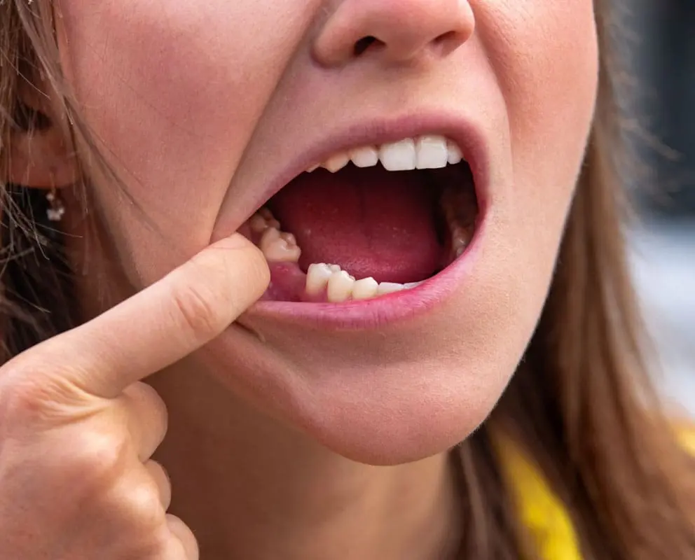
The premature loss, dissolution, or displacement of a tooth extraction clot leads to the painful clinical condition known as alveolar osteitis, commonly called dry socket.
- [ CLOT DISSOLUTION OR DISLODGEMENT ]: Premature fibrinolysis washes away protective plug
- [ ALVEOLAR OSTEITIS (DRY SOCKET) ]: Excruciating throbbing pain radiating to ear and temple
When the protective clot is lost between days two and four, the underlying cortical bone walls and sensory nerves of the jaw lose their biological insulation. Oral bacteria release tissue plasminogen activators that accelerate fibrinolysis, leaving an empty, dry-looking socket devoid of healthy red or white tissue. Patients experience excruciating, radiating pain across the ear, temple, and jaw that does not respond to standard over-the-counter painkillers, accompanied by foul breath and an unpleasant metallic taste. Treatment requires visiting an oral surgeon for socket irrigation, placement of soothing eugenol-impregnated dressings, and targeted pain therapy.
Comprehensive Comparison: Healing Clot vs. Dry Socket vs. Socket Infection
Evaluating clinical symptoms allows patients to distinguish normal granulation tissue from genuine surgical emergencies.
Clinical Parameter | Healthy Healing Clot | Alveolar Osteitis (Dry Socket) | Purulent Socket Infection |
Visual Appearance | Dark red turning to white/cream | Empty, dry socket showing bare gray bone | Swollen red gums, active yellow pus |
Pain Characteristics | Mild to moderate; subsides daily | Excruciating, throbbing, radiating pain | Deep throbbing pain with local swelling |
Pain Onset Timing | Peaks at 24 to 48 hours, then drops | Appears suddenly on days 3 to 5 | Develops after day 4 and steadily worsens |
Tissue Ingrowth | Whitish fibrin and pink granulation | No visible tissue inside socket cavity | Inflamed, friable, bleeding granulation |
Breath and Odor | Normal postoperative taste | Severe fetid odor and foul taste | Distinct bad breath with pus drainage |
Clinical Treatment | Gentle warm saltwater rinses | Sedative medicated alveolar packing | Antibiotics, drainage, and curettage |
Frequently Asked Questions
Why is my tooth extraction clot turning white a few days after surgery?
Your clot is turning white because of normal fibrin deposition and the formation of granulation tissue. As red blood cells break down, a sterile white or cream-colored protein matrix covers the wound to protect the socket while new tissue grows underneath. As long as you have no severe throbbing pain or foul odor, white tissue is a positive sign of healing.
How long does it take for a tooth extraction clot to become secure?
The initial blood clot solidifies within thirty to sixty minutes after surgery, but it remains very fragile for the first seventy-two hours. Between days seven and ten, the clot is fully replaced by stable, anchored granulation tissue that cannot be easily dislodged by normal swallowing or drinking.
What should I do if my extraction socket won't stop bleeding on the first day?
If bleeding persists, fold a sterile gauze pad or a moistened plain black tea bag into a firm square, place it directly over the socket, and bite down with firm, continuous pressure for forty-five minutes while sitting upright. The tannic acid in black tea constricts blood vessels and accelerates clot formation. If heavy, active bleeding continues after two attempts, contact your oral surgeon.
Why is my tooth extraction clot shrinking and creating a dent in my gum?
A shrinking clot is a natural biological step in socket maturation. Fibroblasts and specialized repair cells pull the edges of the healing wound inward to close the opening, while soft tissue slowly fills the space from the base upward. The resulting small indentation will gradually smooth out over several weeks.
Can drinking through a straw really dislodge a wisdom tooth extraction clot?
Yes. Drinking through a straw creates negative air pressure inside your mouth. This suction acts like a vacuum directly over the open extraction socket, pulling the delicate gelatinous clot free from the bone walls and drastically increasing your risk of developing a painful dry socket. Avoid using straws for at least seven to ten days.
About the Author