




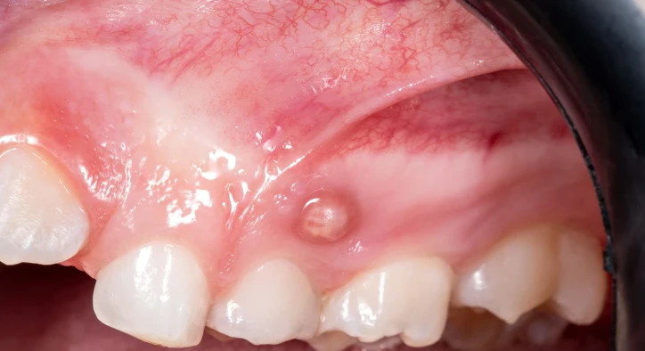

Discovering an unexpected pimple-like bump on the mucosal tissues of the oral cavity often causes immediate concern. Clinically referred to as a parulis or gingival abscess, a gum boil represents an active draining exit point for an underlying chronic infection. Rather than being a localized surface blemish, this oral lesion indicates that bacteria deep within the alveolar bone or periodontal pocket have created an outflow channel to release accumulated purulent fluid into the mouth.
Patients observing an oral lesion frequently ask what is a gum boil and examine the primary gum boil causes to understand where the bacteria originated. A common point of confusion occurs when patients notice a gum boil no pain scenario, mistaking the absence of throbbing ache for a harmless issue. Whether a patient discovers that their gum boil popped while brushing, seeks medical advice on how to get rid of a gum boil, notices a persistent gum boil after tooth extraction, investigates the biological differences in gum boil vs tooth abscess, develops a recurrent gum boil after root canal therapy, or requires definitive gum boil treatment, this clinical guide provides transparent, evidence-based answers.
Table Of Contents
- What Is a Gum Boil and How Does a Parulis Form?
- Etiology: What Are the Primary Gum Boil Causes?
- Symptom Variations: Why Is There Sometimes a Gum Boil No Pain?
- Emergency Response: What Should You Do If a Gum Boil Popped?
- Complications: Extractions, Root Canals, and Differential Diagnosis
- Treatment Options: How to Get Rid of a Gum Boil Permanently
- Comprehensive Comparison: Periapical vs. Periodontal Gum Boils
- Frequently Asked Questions
What Is a Gum Boil and How Does a Parulis Form?
To understand proper management, one must ask: what is a gum boil? A parulis is an elevated mucosal nodule composed of inflammatory granulation tissue that forms at the opening of a chronic fistula tract. When bacteria compromise the dental pulp or periodontal ligament, an infection establishes itself deep inside the jawbone.
- [ DEEP BACTERIAL INFECTION ]: Necrotic dental pulp or deep infected periodontal pocket
- [ PARULIS FORMATION ]: Purulent drainage creates a visible pimple on the gum surface
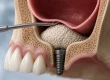
As white blood cells fight the invading pathogens, liquefactive necrosis produces pus. Because pus creates significant hydraulic pressure within enclosed bone spaces, it seeks the path of least resistance. The infection melts away microscopic channels through the cortical plate of the alveolar bone, creates a soft-tissue tunnel called a fistulous tract, and emerges onto the gum surface as an elevated, red or white pustule. This biological pressure release valve discharges small amounts of pus, preventing massive internal pressure buildup.
Etiology: What Are the Primary Gum Boil Causes?
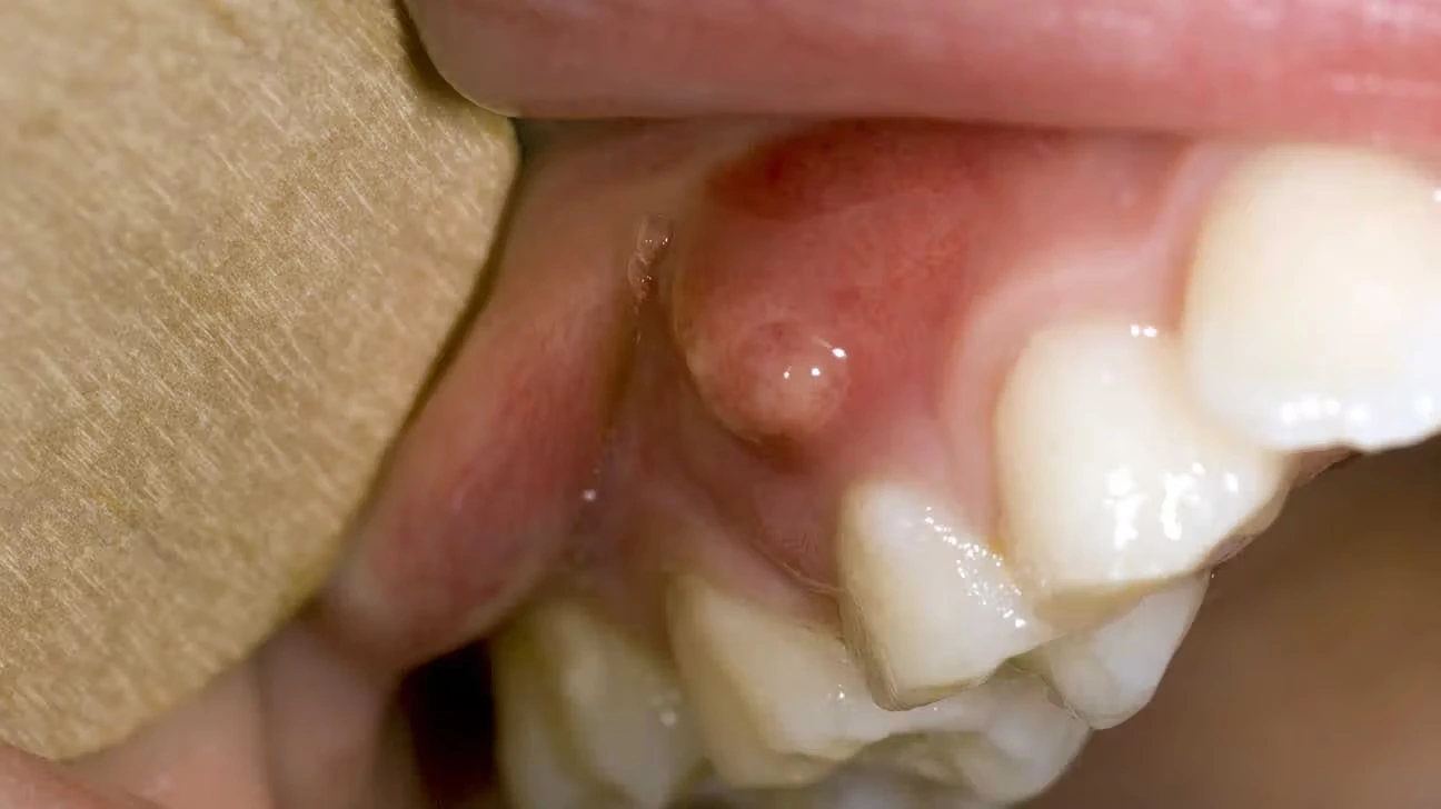
Analyzing gum boil causes requires distinguishing between pulpal and periodontal origins. A boil rarely develops without an underlying infectious source inside the tooth root or gingival architecture.
- Periapical Abscess (Pulp Necrosis): The most common etiology. Untreated deep tooth decay, cracked tooth syndrome, or mechanical dental trauma allows oral bacteria to enter the inner root canal system. Once the nerve tissue dies, bacterial toxins migrate through the apical foramen into the surrounding bone, creating an osteolytic lesion that tunnels outward.
- Periodontal Abscess: Severe, untreated periodontal disease allows deep subgingival calculus and bacterial plaque to accumulate along the root surface. When a deep periodontal pocket becomes blocked, purulent fluid cannot drain coronally, forcing the infection to erupt through the lateral gingiva.
- Foreign Body Impaction: Forcing a popcorn hull, toothbrush bristle, or sharp food fragment subgingivally can introduce pathogens directly into healthy gum tissue, provoking an acute localized abscess.
- Vertical Root Fractures: A microscopic fracture running along the length of an endodontically treated tooth provides an uninterrupted pathway for oral bacteria to colonize the external root surface, forming a stubborn, recurrent fistula.
Understanding these origins is vital when deciding on a definitive, long-term gum boil treatment.
Symptom Variations: Why Is There Sometimes a Gum Boil No Pain?
A major reason patients delay dental appointments is observing a gum boil no pain scenario. In acute infections, internal fluid pressure accumulates within dense bone, compressing sensory nerve fibers and triggering severe, throbbing pain.
- [ CLOSED ABSCESS ]: High fluid pressure inside rigid jawbone causes agonizing, throbbing pain
- [ OPEN PARULIS ]: Fluid decompresses completely, eliminating pain while chronic infection persists
When the infection successfully perforates the bone and forms an open draining parulis, the pressure drops instantly. The absence of pain does not mean the infection has resolved. Instead, it indicates that the infection has transitioned from an acute state to a chronic, low-grade condition. The bacteria continue destroying the supporting alveolar bone silently, releasing purulent drainage and causing bad breath and a sour taste in the mouth without triggering pain receptors.
Emergency Response: What Should You Do If a Gum Boil Popped?
Discovering that your gum boil popped while eating, flossing, or brushing requires prompt, hygienic steps to prevent spreading bacteria across the mouth.
[ PUSTULE RUPTURES ]: Saltwater rinse to cleanse pus, avoid swallowing fluids, maintain hygiene
[ DENTAL VISIT ]: Full clinical debridement, radiographic tracing & root canal/periodontal care
- Rinse with Warm Salt Water: Immediately rinse your mouth gently with half a teaspoon of salt dissolved in warm water. Swish for thirty to sixty seconds and spit out thoroughly to cleanse the mouth of bacteria and soothe inflamed tissues.
- Never Squeeze or Lance the Tissue: Do not attempt to pop, squeeze, or drain a gum boil at home using needles, pins, or fingernails. Prying at the tissue introduces superficial oral bacteria into deep bone structures, causes secondary soft-tissue lacerations, and risks spreading the infection into facial spaces.
- Maintain Gentle Oral Hygiene: Continue brushing and flossing your adjacent natural teeth, but brush very softly over the ruptured lesion to avoid aggravating the raw mucosal opening.
- Schedule an Immediate Evaluation: Remember that spontaneous bursting provides only temporary surface decompression. The underlying infectious factory at the root apex remains fully active, and the boil will refill within days if left unaddressed.
Complications: Extractions, Root Canals, and Differential Diagnosis
Patients frequently encounter unusual healing patterns, requiring clarity regarding gum boil vs tooth abscess dynamics and specialized postoperative scenarios.
- Gum Boil vs Tooth Abscess: A tooth abscess is the primary localized collection of pus within bone or gum tissue, characterized by swelling and throbbing pain when closed. A gum boil (parulis) is the visible external vent or drainage pimple created by an existing tooth abscess. Every gum boil stems from an abscess, but not every abscess develops a gum boil.
- Gum Boil After Tooth Extraction: Developing a gum boil after tooth extraction usually indicates retained root fragments, infected granulation tissue left behind in the socket, a bone sequestrum being rejected, or localized alveolar osteitis complicated by secondary infection.
- Gum Boil After Root Canal: Observing a gum boil after root canal therapy indicates that microscopic bacterial colonies survived within accessory canals, an uncleaned extra canal was missed, apical seals degraded, or an undetected vertical root fracture has developed, requiring endodontic retreatment, apical surgery, or extraction.
Treatment Options: How to Get Rid of a Gum Boil Permanently
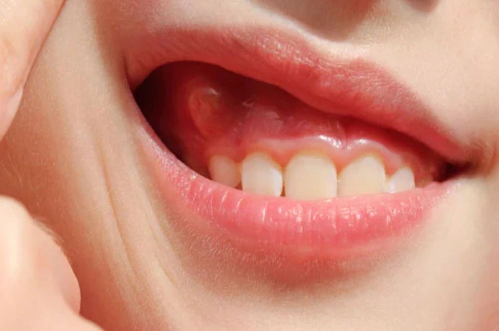
Patients looking for guidance on how to get rid of a gum boil must recognize that home remedies cannot cure the underlying bone pathology. Over-the-counter pain relievers, saltwater rinses, and oral antibiotics may temporarily reduce acute symptoms, but definitive gum boil treatment requires mechanical intervention by a dentist.
[ CLINICAL RADIOGRAPHY ]: Gutta-percha tracing through fistula confirms exact offending tooth root
[ PERMANENT SEALING ]: Dense root canal filling or surgical apicoectomy closes bacterial portal
- Gutta-Percha Fistula Tracing: The dentist inserts a flexible, sterile gutta-percha point into the boil opening and takes an X-ray. The radiopaque point bends along the sinus tract, leading directly to the causative tooth root.
- Non-Surgical Root Canal Therapy: If the boil stems from pulpal necrosis, the clinician disinfects the inner root canal system, removes necrotic tissue, and seals the canals with bioceramic sealers. Once the bacterial reservoir is eliminated, the fistula closes naturally within seven to fourteen days.
- Periodontal Debridement: If the lesion is periodontal, ultrasonic scaling, subgingival curettage, and local antimicrobial irrigation are performed to eliminate subgingival calculus.
- Apicoectomy (Endodontic Microsurgery): If conventional root canal therapy fails to resolve an apical infection, the surgeon resects the root tip, cleans the infected bone cavity, and places a retrograde mineral trioxide aggregate (MTA) seal.
- Tooth Extraction: When a vertical root fracture or extensive bone destruction makes preservation impossible, the tooth is extracted and the socket debrided thoroughly.
Comprehensive Comparison: Periapical vs. Periodontal Gum Boils
Differentiating the clinical characteristics of the two primary forms of gum boils ensures targeted therapy.
Clinical Parameter | Periapical Gum Boil (Endodontic) | Periodontal Gum Boil (Gingival) |
Origin of Infection | Necrotic tooth pulp, root canal failure | Deep periodontal pocket, calculus |
Pulp Vitality Test | Non-vital tooth (no response to cold) | Vital tooth (normal response to cold) |
Location on Gum | Near apical root level, in movable mucosa | Closer to gingival margin, attached gum |
Tooth Mobility | Usually minimal unless bone loss is extensive | Frequently mobile due to ligament loss |
Radiographic Appearance | Radiolucency around the root apex | Horizontal or vertical angular bone loss |
Definitive Treatment | Root canal treatment or apicoectomy | Deep scaling, flap surgery, hygiene |
Frequently Asked Questions
What is a gum boil and will it go away on its own?
A gum boil is a draining pustule on the gum that forms at the exit of a chronic infection originating from a dead tooth nerve or deep periodontal pocket. A gum boil will never go away permanently on its own. While it may shrink or burst temporarily, the infection at the root will continue destroying bone until treated by a dentist.
Why do I have a gum boil no pain in my mouth?
You have no pain because the boil acts as a pressure relief vent. When pus drains out through the fistulous tract into your mouth, internal pressure within the jawbone is eliminated, preventing the severe throbbing ache typical of closed abscesses. However, silent bone destruction continues.
What should I do if my gum boil popped while brushing?
If it pops, gently rinse your mouth with warm salt water to flush away bacteria and soothe the tissues. Do not squeeze or poke the lesion. Contact your dental clinic promptly for an evaluation, as the boil will refill until the root canal infection or periodontal pocket is professionally cleared.
Can antibiotics alone cure a gum boil without a dental procedure?
No. Antibiotics travel through the bloodstream, but necrotic tooth canals have lost their blood supply, preventing the medication from reaching the bacterial source. Antibiotics may temporarily quiet the surrounding soft-tissue swelling, but the boil will return once the prescription ends unless mechanical root canal or periodontal treatment is performed.
Why did I develop a gum boil after root canal treatment?
Developing a boil on an endodontically treated tooth suggests that bacteria survived in microscopic accessory canals, a hidden canal was missed during cleaning, the apical seal failed, or a vertical root fracture has occurred. Your dentist will take digital X-rays to determine whether endodontic retreatment, apical microsurgery, or an extraction is required.
About the Author