




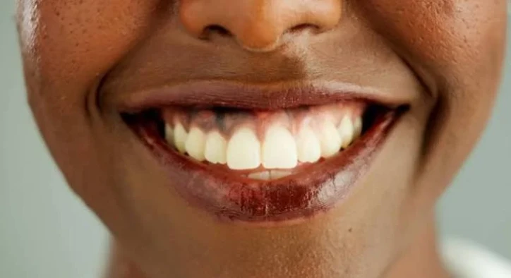

The appearance of dark brown, black, or grey patches along the gingival margins represents a common cosmetic concern known as gingival melanosis or dark gums. When evaluating soft-tissue color variations, understanding gum hyperpigmentation requires examining the biological activity of specialized cells within the basal layer of the oral epithelium. When melanocytes produce an excessive amount of melanin pigment, the naturally pink gum tissue transitions into a spotted or uniformly dark shade, impacting smile harmony even when the underlying teeth are perfectly healthy.
For patients seeking a clear, uniform pink smile line, discovering a reliable gum hyperpigmentation treatment provides a safe, micro-invasive solution. While internet searches often suggest home remedies, true soft-tissue color correction requires targeted clinical intervention to safely reduce melanin density. This comprehensive clinical guide explores the primary biological gum hyperpigmentation causes, details modern treatment for black gums, evaluates protocols for how to get rid of grey gums, showcases expected outcomes of laser gum depigmentation, and provides a structured overview of professional dark gums treatment options.
Table Of Contents
What Is Gum Hyperpigmentation and What Triggers Dark Tissue?
To establish a proper clinical care plan, periodontists must differentiate between natural physiological pigmentation and acquired soft-tissue changes. When defining gum hyperpigmentation, specialists describe an overproduction or localized accumulation of melanin granules produced by melanocytes located in the basal epithelial layer.
The primary gum hyperpigmentation causes include:
- Physiological Melanosis (Genetic Traits): Natural melanin accumulation commonly seen in individuals with darker skin tones, representing a completely healthy genetic variation.
- Smoker’s Melanosis: Chronic exposure to nicotine and polycyclic aromatic hydrocarbons stimulates oral melanocytes to produce excess melanin as a protective response, darkening the anterior gums.
- Heavy Metal & Amalgam Tattoos: Microscopic silver or mercury particles from old dental fillings can embed into the soft tissue, creating localized grey or blue patches. This explains why patients frequently search for how to get rid of grey gums.
- Medication-Induced Pigmentation: Long-term use of specific medications (such as antimalarials, minocycline, or oral contraceptives) can trigger oral tissue discoloration.
- Systemic Conditions: Endocrine disorders like Addison’s disease increase adrenocorticotropic hormone (ACTH), triggering widespread melanin production across the mouth.
- [ HYPERACTIVE MELANOCYTES ]: Excess melanin granules produced in basal epithelial layer
- [ MELANIN GRANULE ACCUMULATION ]: Granules migrate to surface epithelium, darkening gums
- [ DIODE LASER TARGETING ]: Focused light energy selectively targets melanin chromophores
- [ THERMAL ABLATION ]: Hyperactive cells vaporized, allowing clear new tissue to heal
Restorative Strategies: What Is the Best Treatment for Black Gums?
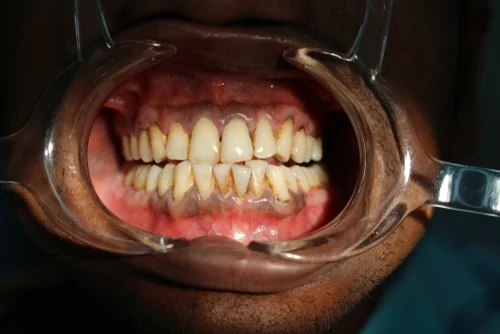
When evaluating treatment for black gums, modern periodontics prioritizes minimally invasive techniques that deliver immediate results with minimal downtime.
1. Laser Gum Depigmentation (Gold Standard)
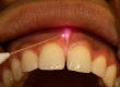
The premier option for dark gums treatment is laser micro-ablation using dental diode or Erbium lasers. The laser light is absorbed selectively by melanin granules, gently vaporizing the hyperpigmented epithelial layer without damaging the underlying connective tissue. This painless procedure requires only mild topical or local anesthesia and heals within a few days to reveal naturally pink tissue.
2. Surgical Scalpel Peeling
A traditional surgical approach where the superficial pigmented layer of the epithelium is carefully abraded using a micro-scalpel. While effective, it involves slightly more post-operative soreness compared to laser therapy.
3. Cryosurgery and Electrosurgery
Utilizing liquid nitrogen freezing or controlled electrical currents to destroy melanin-producing cells. However, these methods offer less depth control than laser therapy, making them less common in modern cosmetic clinics.
Clinical Evaluation: Before and After Gum Depigmentation
Reviewing typical clinical outcomes of laser gum depigmentation demonstrates how removing excess melanin restores natural tissue tone and enhances overall smile aesthetics.
Evaluation Metric | Pre-Treatment Hyperpigmented State | Post-Treatment Laser Restored State |
Gingival Tissue Color | Dark brown, black, or patchy grey spots | Uniform, vibrant, healthy light pink tone |
Melanin Granule Density | Dense melanin concentration in basal layer | Minimal; hyperactive melanocytes ablated |
Post-Operative Healing Time | N/A | 3 to 7 days for complete epithelial renewal |
Patient Comfort Level | Discomfort due to cosmetic self-consciousness | Immediate improvement in smile confidence |
How to Lighten Gums and Maintain Results Long-Term
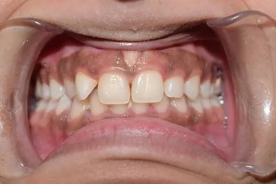
Patients looking for guidance on how to lighten gums permanently must understand that maintaining results requires eliminating external triggers:
- Quit Smoking Immediately: Cessation of tobacco use prevents melanocytes from reactivating and producing new melanin.
- Maintain Excellent Oral Hygiene: Gentle brushing with a soft-bristled brush keeps the tissue healthy and free of inflammatory plaque.
- Replace Old Amalgam Fillings: Upgrading old metal fillings to composite or ceramic prevents new grey amalgam tattoos from forming.
- Schedule Routine Maintenance: Regular periodontal checkups allow your dentist to monitor tissue health and ensure long-term color stability.
Frequently Asked Questions
Is gum hyperpigmentation dangerous to my overall health?
In the vast majority of cases, no. Physiological gum hyperpigmentation is a harmless aesthetic variation. However, if a dark spot appears suddenly, changes shape, bleeds, or grows rapidly, you should see a dentist immediately to rule out rare conditions like oral melanoma.
How long does it take to heal after laser gum depigmentation?
Healing is remarkably fast. The superficial tissue typically heals within 3 to 7 days. Most patients experience minimal discomfort and can resume normal eating and speaking immediately after the procedure.
Can dark gums return after treatment?
If the primary cause was smoking or medication, continuing those habits can cause melanocytes to reactivate over time. However, if external triggers are eliminated, results from laser depigmentation can last for many years or permanently.
Is the treatment for black gums painful?
No. Laser depigmentation is performed under local anesthesia or topical numbing gel, making the procedure virtually painless. Mild soreness for 1 to 2 days after the procedure is easily managed with standard over-the-counter pain relievers.
Can home remedies safely lighten dark gums?
No. Home remedies like baking soda, lemon juice, or abrasive scrubbing cannot reach the basal layer where melanin is produced and can severely burn or damage delicate oral tissues. Professional treatment is the only safe method.
About the Author