





Dental implantology stands as one of the most predictable disciplines in modern reconstructive dentistry, boasting long-term success rates exceeding ninety-five percent. However, when placing fixtures into the posterior mandible, oral surgeons operate in close proximity to critical sensory pathways. Experiencing nerve damage after dental implant surgery represents an uncommon but clinically serious complication that demands immediate identification and targeted management. When an implant fixture encroaches upon the mandibular canal, patient quality of life can be affected by altered sensory perception across the lower lip, chin, and surrounding soft tissues.
Patients experiencing persistent post-operative numbness frequently search for early symptoms of nerve damage after dental implant to determine whether their sensation loss is temporary local anesthesia or acute mechanical trauma. Understanding the timeline of dental implant nerve damage recovery allows clinicians and patients to act within the vital early therapeutic window. Whether dealing with suspected inferior alveolar nerve damage implant impingement, managing a prolonged numb lip after dental implant, evaluating the rare risk of permanent nerve damage from dental implant, or seeking protocols on how to treat nerve damage from dental implant, this clinical guide provides transparent, evidence-based medical insight.
Table Of Contents
- Neuro-Anatomy: Why Does Nerve Injury Occur in the Lower Jaw?
- Symptom Spectrum: What Are the Symptoms of Nerve Damage After Dental Implant?
- Etiology and Classifications of Neural Trauma
- Clinical Diagnostic Workflow and Emergency Management
- Prevention: How Modern Dentistry Eliminates Nerve Damage Risk
- Comparative Matrix: Neuropathy Types and Recovery Prognosis
- Frequently Asked Questions
- How do I know if I have nerve damage after dental implant surgery?
- Is a numb lip after dental implant surgery always permanent?
- What is the critical time window to treat nerve damage from dental implant?
- What should I do if my surgeon dismisses my complaints of numbness?
- How does modern digital dentistry prevent inferior alveolar nerve damage implant complications?
Neuro-Anatomy: Why Does Nerve Injury Occur in the Lower Jaw?
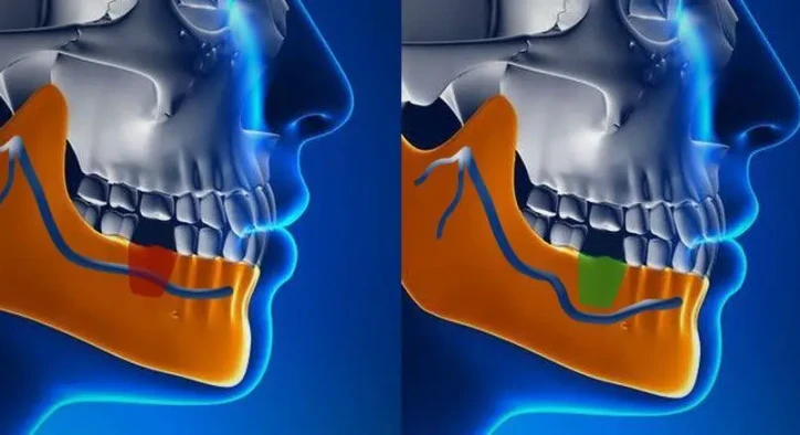
The sensory innervation of the lower third of the face is governed by the mandibular division of the trigeminal nerve. Understanding the precise anatomical path of the inferior alveolar nerve explains why posterior mandibular implant placements carry inherent anatomical risks if three-dimensional planning is bypassed.
- [ INFERIOR ALVEOLAR CANAL ]: Houses sensory nerve bundle beneath mandibular posterior roots
- [ SENSORY NEUROPATHY ]: Mechanical compression, thermal injury, or direct nerve laceration
The inferior alveolar nerve traverses the body of the mandible within a bony conduit called the mandibular canal, supplying sensation to the lower teeth before exiting through the mental foramen near the premolars as the mental nerve, which innervates the lower lip and chin. Because the natural bone ridge resorbs vertically after tooth loss, the distance between the alveolar crest and the nerve canal can shrink considerably. Standard clinical protocols require maintaining a strict safety margin of at least two millimeters between the apex of the implant fixture and the superior border of the mandibular canal. Violating this zone can compress or sever neural fibers.
Symptom Spectrum: What Are the Symptoms of Nerve Damage After Dental Implant?

Recognizing symptoms of nerve damage after dental implant early is essential for preventing permanent sensory loss. While normal local dental anesthesia wears off within three to six hours following surgery, neural injury causes distinct neurological signs.
- Paresthesia (Numbness and Tingling): The most widespread presentation. The patient experiences an absence of sensation or an abnormal pins and needles feeling across the ipsilateral lower lip, chin, labial mucosa, and lower teeth.
- Dysesthesia (Burning and Painful Sensation): An unpleasant, abnormal sensation that feels like spontaneous burning, electrical shocks, stinging, or stabbing pain without any external physical trigger.
- Allodynia (Pain from Non-Painful Stimuli): A condition where light touch, such as shaving, applying makeup, washing the face, or a gentle gust of wind, triggers sharp discomfort.
- Hypoesthesia (Reduced Tactile Sensitivity): Diminished perception of touch, temperature, or pain, often causing patients to bite their lower lip or cheek unknowingly during mastication, or experience unintentional drooling.
When a patient identifies a persistent numb lip after dental implant twenty-four hours after the procedure, immediate diagnostic evaluation is mandatory.
Etiology and Classifications of Neural Trauma
Iatrogenic nerve injuries range from mild physiologic bruising to irreversible structural transection. Oral maxillofacial surgery categorizes these injuries into three distinct classifications established by Sir Herbert Seddon.
[ NEUROPRAXIA ]: Conduction block from compression; nerve fibers intact; resolves in days to weeks
[ AXONOTMESIS ]: Internal axon severed, outer sheath intact; regenerates at 1 mm per day
[ NEUROTMESIS ]: Complete mechanical transection; sheath torn; low chance of spontaneous recovery
- Neuropraxia: The mildest form, caused by mechanical traction, mild compression, or post-surgical edema. The nerve sheath and internal axons remain structurally intact, but nerve conduction is temporarily blocked. Full sensory recovery typically occurs within a few days to six weeks.
- Axonotmesis: Occurs when an implant drill or fixture pinches the nerve, severing internal axons while the surrounding protective epineural sheath remains unbroken. The nerve undergoes Wallerian degeneration and then regenerates from the injury site outward at a rate of approximately one millimeter per day, often recovering over two to six months.
- Neurotmesis: The most severe injury, involving complete anatomical severance or extensive disruption of the nerve trunk caused by direct drill penetration or full fixture invasion. Without prompt microsurgical repair, this condition often results in permanent nerve damage from dental implant.
Clinical Diagnostic Workflow and Emergency Management
When sensory impairment is reported, immediate clinical steps must be initiated. The first thirty-six to forty-eight hours represent the critical window for intervention.
[ CLINICAL COMPLAINT ]: Persistent numbness beyond 24 hours post-implantation
[ EMERGENCY ACTION ]: Immediate fixture back-out (1 to 2 mm) or full removal + high-dose steroids
- Neurosensory Testing: The clinician performs standardized quantitative tests, including light touch detection using cotton fibers, pinprick testing to assess pain receptors, directional stroke testing, and two-point discrimination measurement using a caliper compared against the unaffected side.
- Immediate 3D CBCT Imaging: While two-dimensional panoramic radiographs suffer from vertical magnification distortions, a high-resolution Cone Beam Computed Tomography scan confirms whether the implant apex has breached the cortical roof of the mandibular canal.
- Immediate Surgical Decompression: If imaging demonstrates direct impingement into the nerve canal, the implant must be backed out by one to two millimeters or removed entirely within thirty-six to forty-eight hours. Decompressing the neural tissue early prevents secondary ischemic necrosis and irreversible intraneural scarring.
- Medical Pharmacotherapy: High-dose systemic corticosteroids, such as dexamethasone or prednisolone, are prescribed immediately to reduce intraneural edema. In addition, neuroprotective agents, including high-dose B-complex vitamins, especially methylcobalamin B12, and non-steroidal anti-inflammatory drugs are administered to support peripheral nerve regeneration.
Prevention: How Modern Dentistry Eliminates Nerve Damage Risk
Modern implantology possesses digital diagnostic tools designed to prevent nerve complications. When treated by qualified implantologists, the incidence of permanent neurosensory disturbance drops below one percent.
- Mandibular Canal Tracing via 3D CBCT: Modern implant planning software allows clinicians to digitally highlight the inferior alveolar canal in three-dimensional space, providing cross-sectional measurements with sub-millimeter precision.
- Fully Guided 3D Surgical Stents: Custom surgical guides manufactured via 3D printing control the angulation, depth, and drill stop positioning during bone preparation, preventing the osteotomy bur from penetrating deeper than planned.
- Use of Short and Angled Implants: In cases of severe vertical bone resorption, clinicians utilize extra-short implants (four to six millimeters in length) or angled placements, avoiding the need to operate directly above the nerve pathway.
Comparative Matrix: Neuropathy Types and Recovery Prognosis
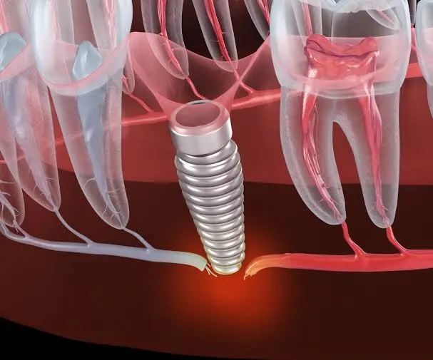
Evaluating the characteristics of sensory disturbances provides insight into long-term functional recovery.
Clinical Parameter | Neuropraxia (Mild Compression) | Axonotmesis (Partial Injury) | Neurotmesis (Complete Severance) |
Primary Cause | Edema, soft-tissue traction | Drill crushing, fixture pressure | Direct drill transection, tear |
Structural Integrity | Epineurium and axon intact | Epineurium intact, axon severed | Complete transection of sheath and axon |
Sensation Quality | Temporary numbness, tingling | Partial numbness, burning sensation | Total loss of touch, temperature, pain |
Recovery Onset | Within 24 to 72 hours | Begins after 3 to 6 weeks | Unlikely without microsurgery |
Full Recovery Time | 2 to 6 weeks | 3 to 9 months | Guarded; permanent deficits likely |
Clinical Action | Steroid therapy, observation | Back-out implant, anti-inflammatories | Immediate removal, nerve exploration |
Frequently Asked Questions
How do I know if I have nerve damage after dental implant surgery?
You may suspect nerve damage if localized numbness in your lower lip, chin, gums, or tongue persists longer than eight to twelve hours after surgery, when local anesthesia should have completely dissipated. The presence of abnormal sensations such as burning, tingling, electrical shocks, or an inability to feel food or liquids on your lip are clear indicators requiring an immediate dental evaluation.
Is a numb lip after dental implant surgery always permanent?
No. In the majority of clinical cases, a numb lip is temporary and caused by neuropraxia, which is minor bruising or swelling around the nerve trunk that resolves within two to eight weeks. Permanent nerve injury is rare and generally occurs only when the nerve bundle is completely severed or if severe implant compression is left untreated for months.
What is the critical time window to treat nerve damage from dental implant?
The most critical time window is the first twenty-four to forty-eight hours post-surgery. If an implant is physically impinging upon the nerve canal, backing the fixture out by one to two millimeters or removing it completely within this initial window drastically increases the chances of full nerve recovery by preventing permanent ischemic damage.
What should I do if my surgeon dismisses my complaints of numbness?
If you experience persistent numbness beyond twenty-four hours and your concerns are dismissed, seek an immediate second opinion from an oral and maxillofacial surgeon or a dental hospital department. Request a high-resolution 3D CBCT scan to independently verify the relationship between the implant apex and your mandibular canal.
How does modern digital dentistry prevent inferior alveolar nerve damage implant complications?
Modern dentistry avoids nerve complications through pre-operative 3D CBCT scans that map the exact location of the mandibular canal down to fractions of a millimeter. Clinicians use virtual planning software and 3D-printed surgical templates equipped with physical drill stops, ensuring the drill cannot penetrate beyond the predetermined two-millimeter safety zone.
About the Author