
Root Canal Treatment is one of the most discussed procedures in modern dentistry, yet it is also among the most misunderstood. When this well known treatment is combined with microscopic technology, it opens a new chapter in precision, visibility, and procedural control. In recent years, microscopic approaches have reshaped how clinicians view and perform Root Canal Treatment, making the process more refined and information driven rather than purely mechanical.
Table Of Contents
- Root Canal Treatment and the Evolution of Dental Visualization
- How Microscopes Are Integrated into Root Canal Treatment
- Scientific Perspective on Precision in Root Canal Treatment
- Root Canal Treatment Under the Microscope and Anatomy Awareness
- Technology, Data, and Root Canal Treatment Outcomes
- Comparing Traditional and Microscopic Root Canal Treatment Approaches
- Root Canal Treatment, Research Trends, and Future Directions
- Analytical Summary of Microscopic Root Canal Treatment
Root Canal Treatment and the Evolution of Dental Visualization
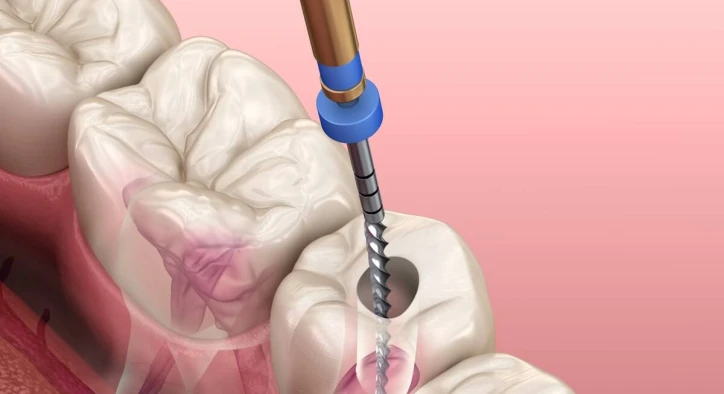
The history of Root Canal Treatment reflects the broader evolution of dentistry itself. Early techniques relied heavily on tactile sensation and basic imaging, which limited how deeply practitioners could observe internal tooth anatomy.
With the introduction of dental microscopes, Root Canal Treatment shifted toward a visibility centered approach. Microscopes offer magnification levels that allow structures inside the tooth to be seen in detail, including micro canals, calcifications, and anatomical variations. This change does not redefine the purpose of Root Canal Treatment, but it reshapes how the procedure is approached from a technical standpoint.
Researchers often describe microscopic Root Canal Treatment as a method that prioritizes observation over assumption. The enhanced field of view supports data driven decision making and aligns with modern minimally invasive philosophies.
How Microscopes Are Integrated into Root Canal Treatment
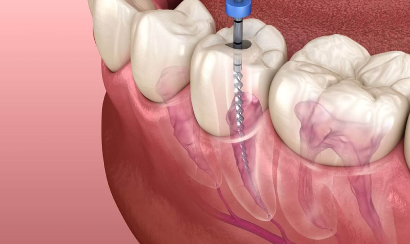
Microscopes are not an add on tool but a central component in microscopic Root Canal Treatment workflows. Their integration begins before any instrumentation and continues throughout the entire procedure.
In a microscopic Root Canal Treatment setting, magnification supports canal identification, debris evaluation, and structural assessment. The microscope remains stationary while the clinician adapts their posture and instrument movement accordingly. This dynamic alters the ergonomics of Root Canal Treatment, potentially influencing how long procedures take and how fatigue is managed.
Studies analyzing microscopic Root Canal Treatment workflows suggest that visual consistency may reduce variability between cases. Rather than relying on generalized anatomical expectations, the procedure becomes case specific and anatomy driven.
Scientific Perspective on Precision in Root Canal Treatment
Precision is a recurring concept in discussions about Root Canal Treatment, particularly when microscopes are involved. Scientific literature often frames precision as the ability to recognize and respond to micro level anatomical details.
In traditional Root Canal Treatment, certain structures may remain unseen due to limitations in magnification. Microscopic Root Canal Treatment challenges this limitation by expanding visual data rather than changing procedural intent. This aligns with broader scientific trends that favor enhanced diagnostics over aggressive intervention.
Researchers emphasize that microscopic Root Canal Treatment does not eliminate complexity. Instead, it reveals it. The increased level of detail requires advanced interpretation skills and deeper anatomical knowledge, reinforcing the role of experience and continuous education.
Root Canal Treatment Under the Microscope and Anatomy Awareness
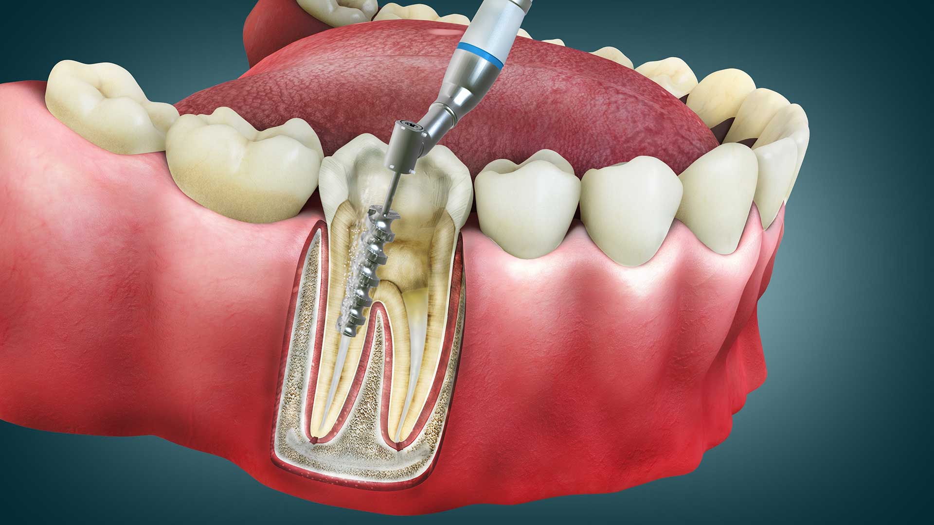
Tooth anatomy is rarely uniform, and Root Canal Treatment often involves navigating variations that are not visible to the naked eye. Microscopic visualization allows these variations to become part of the decision making process rather than unexpected findings.
In microscopic Root Canal Treatment, accessory canals, isthmuses, and internal resorption patterns are more easily identified. This visibility encourages a more analytical approach to internal anatomy and highlights how diverse canal systems can be.
Academic discussions often describe microscopic Root Canal Treatment as anatomy centered rather than technique centered. This distinction matters because it reframes the procedure as an exploration of biological structure rather than a standardized mechanical process.
Technology, Data, and Root Canal Treatment Outcomes
While outcomes are frequently discussed in popular content, scientific discourse around Root Canal Treatment focuses more on data interpretation than promises. Microscopes contribute to this by generating clearer visual data during each stage of the procedure.
In microscopic Root Canal Treatment, visual confirmation becomes part of quality assessment. This includes observing canal cleanliness, structural integrity, and procedural progress. Rather than relying solely on radiographic interpretation, clinicians combine imaging with real time magnified observation.
From a data analysis perspective, microscopic Root Canal Treatment supports documentation and case review. High magnification images can be used for education, peer discussion, and retrospective analysis, contributing to collective professional knowledge.
Comparing Traditional and Microscopic Root Canal Treatment Approaches
Comparisons between traditional and microscopic Root Canal Treatment are common in academic discussions. These comparisons usually focus on visualization, workflow, and interpretive accuracy rather than superiority claims.
Traditional Root Canal Treatment relies on experience and tactile feedback, while microscopic Root Canal Treatment adds a visual dimension to that experience. This combination of sensory input may influence how clinicians assess complexity and procedural limits.
It is important to note that microscopic Root Canal Treatment does not replace foundational endodontic principles. Instead, it builds upon them by offering a clearer view of the same anatomical landscape. This layered approach reflects how technology enhances, rather than replaces, established medical practices.
Root Canal Treatment, Research Trends, and Future Directions
Research into Root Canal Treatment continues to evolve alongside advancements in optical technology. Recent studies explore how magnification affects procedural planning, documentation, and educational outcomes rather than focusing solely on clinical results.
Microscopic Root Canal Treatment is often discussed in relation to training programs. Educational institutions increasingly incorporate magnification early in endodontic education, suggesting a long term shift in how Root Canal Treatment is taught and conceptualized.
Looking forward, researchers speculate that Root Canal Treatment may integrate microscopes with digital imaging, artificial intelligence assisted analysis, and enhanced visualization platforms. These discussions position microscopic Root Canal Treatment as part of a broader ecosystem of data driven dental care.
Analytical Summary of Microscopic Root Canal Treatment
From an analytical standpoint, Root Canal Treatment under the microscope represents a convergence of biology, optics, and procedural science. It does not change the fundamental goals of Root Canal Treatment, but it changes how those goals are pursued and evaluated.
The microscope transforms Root Canal Treatment into a more observational discipline. Each anatomical feature becomes a data point, and each procedural step is guided by enhanced visual feedback. This perspective aligns with modern scientific values that emphasize understanding over assumption.
As dentistry continues to evolve, microscopic Root Canal Treatment stands as an example of how traditional procedures adapt to technological progress without losing their conceptual foundation.
About the Author